 Movie
Movie Controller

[English] 日本語
 Yorodumi
- PDB-4ky4: Crystal structure of non-classical TS inhibitor 2 in complex with... -
+ Open data

- Basic information
| Entry | Database: PDB / ID: 4ky4 | ||||||
|---|---|---|---|---|---|---|---|
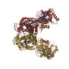
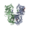
| Title | Crystal structure of non-classical TS inhibitor 2 in complex with Toxoplasma gondii TS-DHFR | ||||||
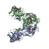
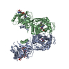
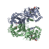
 Components Components | Bifunctional dihydrofolate reductase-thymidylate synthase | ||||||
 Keywords Keywords |  OXIDOREDUCTASE / TRANSFERASE / synthase / bifunctional OXIDOREDUCTASE / TRANSFERASE / synthase / bifunctional | ||||||
| Function / homology |  Function and homology informationthymidylate synthase / thymidylate synthase activity / dTMP biosynthetic process / dihydrofolate reductase / dihydrofolate reductase activity / tetrahydrofolate biosynthetic process / one-carbon metabolic process / methylation Function and homology informationthymidylate synthase / thymidylate synthase activity / dTMP biosynthetic process / dihydrofolate reductase / dihydrofolate reductase activity / tetrahydrofolate biosynthetic process / one-carbon metabolic process / methylationSimilarity search - Function | ||||||
| Biological species |  Toxoplasma gondii (eukaryote) Toxoplasma gondii (eukaryote) | ||||||
| Method | X-RAY DIFFRACTION / SYNCHROTRON / MOLECULAR REPLACEMENT / Resolution: 2.79 Å | ||||||
 Authors Authors | Sharma, H. / Anderson, K.S. | ||||||
 Citation Citation | Journal: ACS Med Chem Lett / Year: 2013 Title: Discovery of potent and selective inhibitors of Toxoplasma gondii thymidylate synthase for opportunistic infections. Authors: Zaware, N. / Sharma, H. / Yang, J. / Devambatla, R.K. / Queener, S.F. / Anderson, K.S. / Gangjee, A. | ||||||
| History |
|
- Structure visualization
| Structure viewer | Molecule: MolmilJmol/JSmol |
|---|
- Downloads & links
-Download
| PDBx/mmCIF format | 4ky4.cif.gz | 804.5 KB | Display | PDBx/mmCIF format |
|---|---|---|---|---|
| PDB format | pdb4ky4.ent.gz | 664.3 KB | Display | PDB format |
| PDBx/mmJSON format | 4ky4.json.gz | Tree view | PDBx/mmJSON format | |
| Others |  Other downloads Other downloads |
-Validation report
| Arichive directory | https://data.pdbj.org/pub/pdb/validation_reports/ky/4ky4ftp://data.pdbj.org/pub/pdb/validation_reports/ky/4ky4 | HTTPS FTP |
|---|
-Related structure data
| Related structure data |  4kyaC  4eilS S: Starting model for refinement C: citing same article ( |
|---|---|
| Similar structure data |

-Links
 PDBj
PDBj

- Assembly
| Deposited unit | 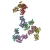
| ||||||||
|---|---|---|---|---|---|---|---|---|---|
| 1 | 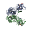
| ||||||||
| 2 | 
| ||||||||
| 3 | 
| ||||||||
| 4 | 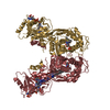
| ||||||||
| Unit cell |
|
-Components
| #1: Protein | Mass: 68842.656 Da / Num. of mol.: 8 Source method: isolated from a genetically manipulated source Source: (gene. exp.) Toxoplasma gondii (eukaryote) / Production host:  Escherichia coli (E. coli) Escherichia coli (E. coli)References: UniProt: Q07422, dihydrofolate reductase, thymidylate synthase#2: Chemical | ChemComp-UMP / Deoxyuridine monophosphate  Mass: 308.182 Da / Num. of mol.: 8 / Source method: obtained synthetically / Formula: C9H13N2O8P Mass: 308.182 Da / Num. of mol.: 8 / Source method: obtained synthetically / Formula: C9H13N2O8P#3: Chemical | ChemComp-04J / Aminopterin  Mass: 440.413 Da / Num. of mol.: 8 / Source method: obtained synthetically / Formula: C19H20N8O5 / Comment: chemotherapy, antineoplastic*YM Mass: 440.413 Da / Num. of mol.: 8 / Source method: obtained synthetically / Formula: C19H20N8O5 / Comment: chemotherapy, antineoplastic*YM#4: Chemical | ChemComp-NDP / Nicotinamide adenine dinucleotide phosphate  Mass: 745.421 Da / Num. of mol.: 8 / Source method: obtained synthetically / Formula: C21H30N7O17P3 Mass: 745.421 Da / Num. of mol.: 8 / Source method: obtained synthetically / Formula: C21H30N7O17P3#5: Chemical | ChemComp-1UE /   Mass: 308.358 Da / Num. of mol.: 8 / Source method: obtained synthetically / Formula: C16H12N4OS Mass: 308.358 Da / Num. of mol.: 8 / Source method: obtained synthetically / Formula: C16H12N4OS |
|---|
-Experimental details
-Experiment
| Experiment | Method: X-RAY DIFFRACTION / Number of used crystals: 1 |
|---|
- Sample preparation
| Crystal | Density Matthews: 2.49 Å3/Da / Density % sol: 50.51 % |
|---|---|
| Crystal grow | Temperature: 293 K / Method: vapor diffusion, hanging drop / pH: 7.3 Details: 18% PEG 3350, 0.1 M Potassium Formate, pH 7.3, VAPOR DIFFUSION, HANGING DROP, temperature 293K |
-Data collection
| Diffraction |
| |||||||||||||||
|---|---|---|---|---|---|---|---|---|---|---|---|---|---|---|---|---|
| Diffraction source | Source: SYNCHROTRON / Site: NSLS  / Beamline: X25 / Wavelength: 1.1 Å / Beamline: X25 / Wavelength: 1.1 Å | |||||||||||||||
| Detector | Type: PSI PILATUS 6M / Detector: PIXEL / Date: Nov 4, 2012 | |||||||||||||||
| Radiation |
| |||||||||||||||
| Radiation wavelength | Wavelength: 1.1 Å / Relative weight: 1 | |||||||||||||||
| Reflection | Resolution: 2.79→48.28 Å / Num. all: 128608 / Num. obs: 121332 / % possible obs: 94.34 % / Observed criterion σ(F): 2 / Observed criterion σ(I): 2 |
- Processing
| Software |
| |||||||||||||||||||||||||||||||||||||||||||||||||||||||||||||||||||||||||||
|---|---|---|---|---|---|---|---|---|---|---|---|---|---|---|---|---|---|---|---|---|---|---|---|---|---|---|---|---|---|---|---|---|---|---|---|---|---|---|---|---|---|---|---|---|---|---|---|---|---|---|---|---|---|---|---|---|---|---|---|---|---|---|---|---|---|---|---|---|---|---|---|---|---|---|---|---|
| Refinement | Method to determine structure: MOLECULAR REPLACEMENT Starting model: PDB ENTRY 4EIL Resolution: 2.79→48.28 Å / Cor.coef. Fo:Fc: 0.934 / Cor.coef. Fo:Fc free: 0.905 / Occupancy max: 1 / Occupancy min: 0.42 / SU B: 16.948 / SU ML: 0.33 / Cross valid method: THROUGHOUT / σ(F): 2 / ESU R Free: 0.395 Stereochemistry target values: MAXIMUM LIKELIHOOD WITH PHASES Details: HYDROGENS HAVE BEEN ADDED IN THE RIDING POSITIONS U VALUES: REFINED INDIVIDUALLY
| |||||||||||||||||||||||||||||||||||||||||||||||||||||||||||||||||||||||||||
| Solvent computation | Ion probe radii: 0.8 Å / Shrinkage radii: 0.8 Å / VDW probe radii: 1.2 Å / Solvent model: MASK | |||||||||||||||||||||||||||||||||||||||||||||||||||||||||||||||||||||||||||
| Displacement parameters | Biso max: 202.23 Å2 / Biso mean: 79.025 Å2 / Biso min: 42.59 Å2
| |||||||||||||||||||||||||||||||||||||||||||||||||||||||||||||||||||||||||||
| Refinement step | Cycle: LAST / Resolution: 2.79→48.28 Å
| |||||||||||||||||||||||||||||||||||||||||||||||||||||||||||||||||||||||||||
| Refine LS restraints |
| |||||||||||||||||||||||||||||||||||||||||||||||||||||||||||||||||||||||||||
| LS refinement shell | Resolution: 2.793→2.866 Å / Total num. of bins used: 20
|
