 Movie
Movie Controller

[English] 日本語
 Yorodumi
- PDB-4kwu: 1.9 Angstrom resolution crystal structure of uncharacterized prot... -
+ Open data

- Basic information
| Entry | Database: PDB / ID: 4kwu | ||||||
|---|---|---|---|---|---|---|---|
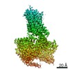
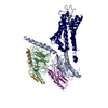
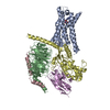
| Title | 1.9 Angstrom resolution crystal structure of uncharacterized protein lmo2446 from Listeria monocytogenes EGD-e in complex with alpha-D-glucose, beta-D-glucose, magnesium and calcium | ||||||
 Components Components | Lmo2446 protein | ||||||
 Keywords Keywords |  STRUCTURAL GENOMICS / UNKNOWN FUNCTION / Center for Structural Genomics of Infectious Diseases / CSGID / NIAID / National Institute of Allergy and Infectious Diseases / TIM-barrel / beta-fold STRUCTURAL GENOMICS / UNKNOWN FUNCTION / Center for Structural Genomics of Infectious Diseases / CSGID / NIAID / National Institute of Allergy and Infectious Diseases / TIM-barrel / beta-fold | ||||||
| Function / homology |  Function and homology information Function and homology informationhydrolase activity, hydrolyzing O-glycosyl compounds / carbohydrate binding / carbohydrate metabolic process / metal ion bindingSimilarity search - Function | ||||||
| Biological species |  Listeria monocytogenes (bacteria) Listeria monocytogenes (bacteria) | ||||||
| Method | X-RAY DIFFRACTION / SYNCHROTRON / MOLECULAR REPLACEMENT / Resolution: 1.9 Å | ||||||
 Authors Authors | Halavaty, A.S. / Minasov, G. / Dubrovska, I. / Winsor, J. / Shuvalova, L. / Peterson, S. / Anderson, W.F. / Center for Structural Genomics of Infectious Diseases (CSGID) | ||||||
 Citation Citation | Journal: To be Published Title: 1.9 Angstrom resolution crystal structure of uncharacterized protein lmo2446 from Listeria monocytogenes EGD-e in complex with alpha-D-glucose, beta-D-glucose, magnesium and calcium Authors: Halavaty, A.S. / Minasov, G. / Dubrovska, I. / Winsor, J. / Shuvalova, L. / Peterson, S. / Anderson, W.F. / Center for Structural Genomics of Infectious Diseases (CSGID) | ||||||
| History |
|
- Structure visualization
| Structure viewer | Molecule: MolmilJmol/JSmol |
|---|
- Downloads & links
-Download
| PDBx/mmCIF format | 4kwu.cif.gz | 487.2 KB | Display | PDBx/mmCIF format |
|---|---|---|---|---|
| PDB format | pdb4kwu.ent.gz | 397.5 KB | Display | PDB format |
| PDBx/mmJSON format | 4kwu.json.gz | Tree view | PDBx/mmJSON format | |
| Others |  Other downloads Other downloads |
-Validation report
| Arichive directory | https://data.pdbj.org/pub/pdb/validation_reports/kw/4kwuftp://data.pdbj.org/pub/pdb/validation_reports/kw/4kwu | HTTPS FTP |
|---|
-Related structure data
| Related structure data |  4kmqS S: Starting model for refinement |
|---|---|
| Similar structure data | |
| Other databases |


-Links
 PDBj
PDBj


- Assembly
| Deposited unit | 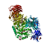
| ||||||||
|---|---|---|---|---|---|---|---|---|---|
| 1 |
| ||||||||
| Unit cell |
|
-Components
-Protein , 1 types, 1 molecules A
| #1: Protein | Mass: 122520.227 Da / Num. of mol.: 1 / Fragment: UNP residues 31-1091 Source method: isolated from a genetically manipulated source Source: (gene. exp.) Listeria monocytogenes (bacteria) / Strain: EGD-e / Gene: lmo2446 / Plasmid: pMCSG7 / Production host: Escherichia coli (E. coli) / Strain (production host): BL21-Magic / References: UniProt: Q8Y4J2 |
|---|
-Sugars , 2 types, 10 molecules 

| #4: Sugar | ChemComp-BGC / Glucose Type: D-saccharide, beta linking / Mass: 180.156 Da / Num. of mol.: 4 Type: D-saccharide, beta linking / Mass: 180.156 Da / Num. of mol.: 4Source method: isolated from a genetically manipulated source Formula: C6H12O6 #5: Sugar | ChemComp-GLC / Glucose Type: D-saccharide, alpha linking / Mass: 180.156 Da / Num. of mol.: 6 Type: D-saccharide, alpha linking / Mass: 180.156 Da / Num. of mol.: 6Source method: isolated from a genetically manipulated source Formula: C6H12O6 |
|---|
-Non-polymers , 3 types, 1241 molecules 


| #2: Chemical | ChemComp-MG /  Mass: 24.305 Da / Num. of mol.: 8 / Source method: obtained synthetically / Formula: Mg Mass: 24.305 Da / Num. of mol.: 8 / Source method: obtained synthetically / Formula: Mg#3: Chemical | ChemComp-CA / |  Mass: 40.078 Da / Num. of mol.: 1 / Source method: obtained synthetically / Formula: Ca Mass: 40.078 Da / Num. of mol.: 1 / Source method: obtained synthetically / Formula: Ca#6: Water | ChemComp-HOH / | WaterMass: 18.015 Da / Num. of mol.: 1232 / Source method: isolated from a natural source / Formula: H2O |
|---|
-Experimental details
-Experiment
| Experiment | Method: X-RAY DIFFRACTION / Number of used crystals: 1 |
|---|
- Sample preparation
| Crystal | Density Matthews: 2.53 Å3/Da / Density % sol: 51.37 % |
|---|---|
| Crystal grow | Temperature: 295 K / Method: vapor diffusion, sitting drop / pH: 8.3 Details: crystallization conditions - 0.2 M magnesium formate, 20% (w/v) PEG3350 (The JCSG+ suite: A5), protein - 7.3 mg/mL in 10 mM Tris-HCl pH8.3 500 mM NaCl 5 mM BME, soak/cryo - crystallization ...Details: crystallization conditions - 0.2 M magnesium formate, 20% (w/v) PEG3350 (The JCSG+ suite: A5), protein - 7.3 mg/mL in 10 mM Tris-HCl pH8.3 500 mM NaCl 5 mM BME, soak/cryo - crystallization conditions + 50% glucose, VAPOR DIFFUSION, SITTING DROP, temperature 295K |
-Data collection
| Diffraction | Mean temperature: 100 K |
|---|---|
| Diffraction source | Source: SYNCHROTRON / Site: APS  / Beamline: 21-ID-D / Wavelength: 1.0782 Å / Beamline: 21-ID-D / Wavelength: 1.0782 Å |
| Detector | Type: MARMOSAIC 300 mm CCD / Detector: CCD / Date: Mar 28, 2013 / Details: Mirror |
| Radiation | Monochromator: Si(111)Channel / Protocol: SINGLE WAVELENGTH / Monochromatic (M) / Laue (L): M / Scattering type: x-ray |
| Radiation wavelength | Wavelength: 1.0782 Å / Relative weight: 1 |
| Reflection | Resolution: 1.9→30 Å / Num. all: 94351 / Num. obs: 94351 / % possible obs: 98.5 % / Observed criterion σ(I): -3 / Redundancy: 3.6 % / Biso Wilson estimate: 22.7 Å2 / Rmerge(I) obs: 0.076 / Net I/σ(I): 15.8 |
| Reflection shell | Resolution: 1.9→1.93 Å / Redundancy: 3.6 % / Rmerge(I) obs: 0.568 / Mean I/σ(I) obs: 2.6 / Num. unique all: 4671 / % possible all: 97.5 |
- Processing
| Software |
| |||||||||||||||||||||||||||||||||||||||||||||||||||||||||||||||||||||||||||||||||||||||||||||||||||||||||||||||||||||||||||||||||||||||||||||||||||||||||||||||||||||||||||||||
|---|---|---|---|---|---|---|---|---|---|---|---|---|---|---|---|---|---|---|---|---|---|---|---|---|---|---|---|---|---|---|---|---|---|---|---|---|---|---|---|---|---|---|---|---|---|---|---|---|---|---|---|---|---|---|---|---|---|---|---|---|---|---|---|---|---|---|---|---|---|---|---|---|---|---|---|---|---|---|---|---|---|---|---|---|---|---|---|---|---|---|---|---|---|---|---|---|---|---|---|---|---|---|---|---|---|---|---|---|---|---|---|---|---|---|---|---|---|---|---|---|---|---|---|---|---|---|---|---|---|---|---|---|---|---|---|---|---|---|---|---|---|---|---|---|---|---|---|---|---|---|---|---|---|---|---|---|---|---|---|---|---|---|---|---|---|---|---|---|---|---|---|---|---|---|---|---|
| Refinement | Method to determine structure: MOLECULAR REPLACEMENT Starting model: 4KMQ Resolution: 1.9→29.92 Å / Cor.coef. Fo:Fc: 0.977 / Cor.coef. Fo:Fc free: 0.963 / SU B: 5.408 / SU ML: 0.072 / Isotropic thermal model: Isotropic / Cross valid method: THROUGHOUT / ESU R: 0.117 / ESU R Free: 0.11 / Stereochemistry target values: MAXIMUM LIKELIHOOD / Details: HYDROGENS HAVE BEEN ADDED IN THE RIDING POSITIONS
| |||||||||||||||||||||||||||||||||||||||||||||||||||||||||||||||||||||||||||||||||||||||||||||||||||||||||||||||||||||||||||||||||||||||||||||||||||||||||||||||||||||||||||||||
| Solvent computation | Ion probe radii: 0.8 Å / Shrinkage radii: 0.8 Å / VDW probe radii: 1.2 Å / Solvent model: BABINET MODEL WITH MASK | |||||||||||||||||||||||||||||||||||||||||||||||||||||||||||||||||||||||||||||||||||||||||||||||||||||||||||||||||||||||||||||||||||||||||||||||||||||||||||||||||||||||||||||||
| Displacement parameters | Biso mean: 20.468 Å2
| |||||||||||||||||||||||||||||||||||||||||||||||||||||||||||||||||||||||||||||||||||||||||||||||||||||||||||||||||||||||||||||||||||||||||||||||||||||||||||||||||||||||||||||||
| Refinement step | Cycle: LAST / Resolution: 1.9→29.92 Å
| |||||||||||||||||||||||||||||||||||||||||||||||||||||||||||||||||||||||||||||||||||||||||||||||||||||||||||||||||||||||||||||||||||||||||||||||||||||||||||||||||||||||||||||||
| Refine LS restraints |
| |||||||||||||||||||||||||||||||||||||||||||||||||||||||||||||||||||||||||||||||||||||||||||||||||||||||||||||||||||||||||||||||||||||||||||||||||||||||||||||||||||||||||||||||
| LS refinement shell | Resolution: 1.9→1.949 Å / Total num. of bins used: 20
| |||||||||||||||||||||||||||||||||||||||||||||||||||||||||||||||||||||||||||||||||||||||||||||||||||||||||||||||||||||||||||||||||||||||||||||||||||||||||||||||||||||||||||||||
| Refinement TLS params. | Method: refined / Refine-ID: X-RAY DIFFRACTION
| |||||||||||||||||||||||||||||||||||||||||||||||||||||||||||||||||||||||||||||||||||||||||||||||||||||||||||||||||||||||||||||||||||||||||||||||||||||||||||||||||||||||||||||||
| Refinement TLS group |
|
