 Movie
Movie Controller


+ Open data

- Basic information
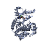
| Entry | Database: PDB / ID: 4ks8 | ||||||
|---|---|---|---|---|---|---|---|
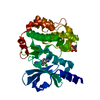
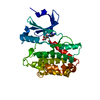
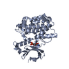
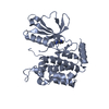
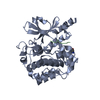
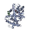
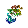
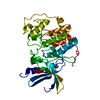
| Title | PAK6 kinase domain in complex with sunitinib | ||||||
 Components Components | Serine/threonine-protein kinase PAK 6 | ||||||
 Keywords Keywords | TRANSFERASE/TRANSFERASE INHIBITOR /  Protein Kinase / Phosphotransfer / Phosphorylation / TRANSFERASE-TRANSFERASE INHIBITOR complex Protein Kinase / Phosphotransfer / Phosphorylation / TRANSFERASE-TRANSFERASE INHIBITOR complex | ||||||
| Function / homology |  Function and homology information Function and homology informationneuron projection arborization / RHOD GTPase cycle / Activation of RAC1 / neuron projection extension / RHOV GTPase cycle / regulation of MAPK cascade / RHOH GTPase cycle / CDC42 GTPase cycle / cytoskeleton organization / RAC1 GTPase cycle ...neuron projection arborization / RHOD GTPase cycle / Activation of RAC1 / neuron projection extension / RHOV GTPase cycle / regulation of MAPK cascade / RHOH GTPase cycle / CDC42 GTPase cycle / cytoskeleton organization / RAC1 GTPase cycle / locomotory behavior / learning / memory / fibrillar center / cell junction / postsynaptic density / non-specific serine/threonine protein kinase / intracellular signal transduction / cadherin binding / phosphorylation / protein serine kinase activity / protein serine/threonine kinase activity / apoptotic process / regulation of DNA-templated transcription / nucleoplasm / ATP binding / cytosol / cytoplasmSimilarity search - Function | ||||||
| Biological species |  Homo sapiens (human) Homo sapiens (human) | ||||||
| Method | X-RAY DIFFRACTION / SYNCHROTRON / MOLECULAR REPLACEMENT / Resolution: 1.95 Å | ||||||
 Authors Authors | Gao, J. / Boggon, T.J. | ||||||
 Citation Citation | Journal: Plos One / Year: 2013 Title: Substrate and Inhibitor Specificity of the Type II p21-Activated Kinase, PAK6. Authors: Gao, J. / Ha, B.H. / Lou, H.J. / Morse, E.M. / Zhang, R. / Calderwood, D.A. / Turk, B.E. / Boggon, T.J. | ||||||
| History |
|
- Structure visualization
| Structure viewer | Molecule: MolmilJmol/JSmol |
|---|
- Downloads & links
-Download
| PDBx/mmCIF format | 4ks8.cif.gz | 75.4 KB | Display | PDBx/mmCIF format |
|---|---|---|---|---|
| PDB format | pdb4ks8.ent.gz | 54.7 KB | Display | PDB format |
| PDBx/mmJSON format | 4ks8.json.gz | Tree view | PDBx/mmJSON format | |
| Others |  Other downloads Other downloads |
-Validation report
| Arichive directory | https://data.pdbj.org/pub/pdb/validation_reports/ks/4ks8ftp://data.pdbj.org/pub/pdb/validation_reports/ks/4ks8 | HTTPS FTP |
|---|
-Related structure data
| Related structure data |  4ks7SC S: Starting model for refinement C: citing same article ( |
|---|---|
| Similar structure data |


-Links
 PDBj
PDBj


- Assembly
| Deposited unit | 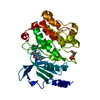
| ||||||||
|---|---|---|---|---|---|---|---|---|---|
| 1 |
| ||||||||
| Unit cell |
|
-Components
| #1: Protein | Mass: 33301.551 Da / Num. of mol.: 1 / Fragment: Kinase domain Source method: isolated from a genetically manipulated source Source: (gene. exp.) Homo sapiens (human) / Gene: PAK6, PAK5 / Plasmid: pET-32 / Production host:  Escherichia coli (E. coli) Escherichia coli (E. coli)References: UniProt: Q9NQU5, non-specific serine/threonine protein kinase |
|---|---|
| #2: Chemical | ChemComp-B49 / Sunitinib  Mass: 398.474 Da / Num. of mol.: 1 / Source method: obtained synthetically / Formula: C22H27FN4O2 / Comment: medication, anticancer, inhibitor*YM Mass: 398.474 Da / Num. of mol.: 1 / Source method: obtained synthetically / Formula: C22H27FN4O2 / Comment: medication, anticancer, inhibitor*YM |
| #3: Water | ChemComp-HOH / Water Mass: 18.015 Da / Num. of mol.: 171 / Source method: isolated from a natural source / Formula: H2O Mass: 18.015 Da / Num. of mol.: 171 / Source method: isolated from a natural source / Formula: H2O |
-Experimental details
-Experiment
| Experiment | Method: X-RAY DIFFRACTION / Number of used crystals: 1 |
|---|
- Sample preparation
| Crystal | Density Matthews: 2.24 Å3/Da / Density % sol: 45.04 % |
|---|---|
| Crystal grow | Temperature: 277 K / Method: vapor diffusion, sitting drop / pH: 8 Details: 30% (v/v) isopropanol, 0.1M MES, pH 8.0, VAPOR DIFFUSION, SITTING DROP, temperature 277K |
-Data collection
| Diffraction | Mean temperature: 100 K |
|---|---|
| Diffraction source | Source: SYNCHROTRON / Site: NSLS  / Beamline: X6A / Wavelength: 1 Å / Beamline: X6A / Wavelength: 1 Å |
| Detector | Type: ADSC QUANTUM 270 / Detector: CCD / Date: Nov 18, 2012 / Details: mirrors |
| Radiation | Monochromator: mirrors / Protocol: SINGLE WAVELENGTH / Monochromatic (M) / Laue (L): M / Scattering type: x-ray |
| Radiation wavelength | Wavelength: 1 Å / Relative weight: 1 |
| Reflection | Resolution: 1.95→50 Å / Num. obs: 22562 / % possible obs: 100 % / Observed criterion σ(F): 0 / Observed criterion σ(I): 0 / Redundancy: 6.8 % / Biso Wilson estimate: 20 Å2 / Rsym value: 0.117 / Net I/σ(I): 17.47 |
| Reflection shell | Resolution: 1.95→2.02 Å / Redundancy: 6.7 % / Mean I/σ(I) obs: 2 / Num. unique all: 2187 / Rsym value: 0.885 / % possible all: 100 |
- Processing
| Software |
| ||||||||||||||||||||||||||||||||||||||||||||||||||||||||||||||||||||||||||||||||||||||||||||||||||||||||||||||||||||||||||||||||||||||||||||||||||||||||||||||||||||||||||
|---|---|---|---|---|---|---|---|---|---|---|---|---|---|---|---|---|---|---|---|---|---|---|---|---|---|---|---|---|---|---|---|---|---|---|---|---|---|---|---|---|---|---|---|---|---|---|---|---|---|---|---|---|---|---|---|---|---|---|---|---|---|---|---|---|---|---|---|---|---|---|---|---|---|---|---|---|---|---|---|---|---|---|---|---|---|---|---|---|---|---|---|---|---|---|---|---|---|---|---|---|---|---|---|---|---|---|---|---|---|---|---|---|---|---|---|---|---|---|---|---|---|---|---|---|---|---|---|---|---|---|---|---|---|---|---|---|---|---|---|---|---|---|---|---|---|---|---|---|---|---|---|---|---|---|---|---|---|---|---|---|---|---|---|---|---|---|---|---|---|---|---|
| Refinement | Method to determine structure: MOLECULAR REPLACEMENT Starting model: PDB entry 4KS7 Resolution: 1.95→38.46 Å / Cor.coef. Fo:Fc: 0.94 / Cor.coef. Fo:Fc free: 0.91 / SU B: 4.357 / SU ML: 0.124 / Cross valid method: THROUGHOUT / ESU R: 0.203 / ESU R Free: 0.177 / Stereochemistry target values: MAXIMUM LIKELIHOOD / Details: HYDROGENS HAVE BEEN ADDED IN THE RIDING POSITIONS
| ||||||||||||||||||||||||||||||||||||||||||||||||||||||||||||||||||||||||||||||||||||||||||||||||||||||||||||||||||||||||||||||||||||||||||||||||||||||||||||||||||||||||||
| Solvent computation | Ion probe radii: 0.8 Å / Shrinkage radii: 0.8 Å / VDW probe radii: 1.2 Å / Solvent model: MASK | ||||||||||||||||||||||||||||||||||||||||||||||||||||||||||||||||||||||||||||||||||||||||||||||||||||||||||||||||||||||||||||||||||||||||||||||||||||||||||||||||||||||||||
| Displacement parameters | Biso mean: 28.004 Å2
| ||||||||||||||||||||||||||||||||||||||||||||||||||||||||||||||||||||||||||||||||||||||||||||||||||||||||||||||||||||||||||||||||||||||||||||||||||||||||||||||||||||||||||
| Refinement step | Cycle: LAST / Resolution: 1.95→38.46 Å
| ||||||||||||||||||||||||||||||||||||||||||||||||||||||||||||||||||||||||||||||||||||||||||||||||||||||||||||||||||||||||||||||||||||||||||||||||||||||||||||||||||||||||||
| Refine LS restraints |
| ||||||||||||||||||||||||||||||||||||||||||||||||||||||||||||||||||||||||||||||||||||||||||||||||||||||||||||||||||||||||||||||||||||||||||||||||||||||||||||||||||||||||||
| LS refinement shell | Resolution: 1.95→1.997 Å / Total num. of bins used: 20
|
