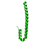
 Movie
Movie Controller

[English] 日本語
 Yorodumi
- PDB-4kmc: Structure analysis of M. Tuberculosis rRNA transcriptional regula... -
+ Open data

- Basic information
| Entry | Database: PDB / ID: 4kmc | ||||||
|---|---|---|---|---|---|---|---|
| Title | Structure analysis of M. Tuberculosis rRNA transcriptional regulator CarD and its interaction with T. Aquaticus RNA polymerase-BETA1 domain | ||||||
 Components Components | RNA polymerase-binding transcription factor CarD | ||||||
 Keywords Keywords |  TRANSCRIPTION / UNKNOWN FOLD / rRNA TRANSCRIPTIONAL REGULATOR / RNA POLYMERASE BETA1 DOMAIN TRANSCRIPTION / UNKNOWN FOLD / rRNA TRANSCRIPTIONAL REGULATOR / RNA POLYMERASE BETA1 DOMAIN | ||||||
| Function / homology |  Function and homology information Function and homology information | ||||||
| Biological species |   Mycobacterium tuberculosis (bacteria) Mycobacterium tuberculosis (bacteria) | ||||||
| Method | X-RAY DIFFRACTION / SYNCHROTRON / SAD / Resolution: 2.15 Å | ||||||
 Authors Authors | Gangwar, S.P. / Meena, S.R. / Saxena, A.K. | ||||||
 Citation Citation | Journal: Acta Crystallogr.,Sect.F / Year: 2014 Title: Structure of the carboxy-terminal domain of Mycobacterium tuberculosis CarD protein: an essential rRNA transcriptional regulator. Authors: Gangwar, S.P. / Meena, S.R. / Saxena, A.K. | ||||||
| History |
|
- Structure visualization
| Structure viewer | Molecule: MolmilJmol/JSmol |
|---|
- Downloads & links
-Download
| PDBx/mmCIF format | 4kmc.cif.gz | 42.2 KB | Display | PDBx/mmCIF format |
|---|---|---|---|---|
| PDB format | pdb4kmc.ent.gz | 30.3 KB | Display | PDB format |
| PDBx/mmJSON format | 4kmc.json.gz | Tree view | PDBx/mmJSON format | |
| Others |  Other downloads Other downloads |
-Validation report
| Arichive directory | https://data.pdbj.org/pub/pdb/validation_reports/km/4kmcftp://data.pdbj.org/pub/pdb/validation_reports/km/4kmc | HTTPS FTP |
|---|
-Related structure data
| Similar structure data |
|---|


-Links
 PDBj
PDBj- Assembly
| Deposited unit | 
| ||||||||
|---|---|---|---|---|---|---|---|---|---|
| 1 |
| ||||||||
| 2 | 
| ||||||||
| Unit cell |
| ||||||||
| Components on special symmetry positions |
|
-Components
| #1: Protein | Mass: 8883.027 Da / Num. of mol.: 1 / Fragment: C terminal domain, UNP residues 83-162 Source method: isolated from a genetically manipulated source Source: (gene. exp.) Mycobacterium tuberculosis (bacteria) / Strain: H37Rv / Gene: carD, MT3689, Rv3583c / Plasmid: pET28a(+) / Production host: Escherichia coli (E. coli) / Strain (production host): BL21(DE3) / References: UniProt: O53568, UniProt: P9WJG3*PLUS |
|---|---|
| #2: Water | ChemComp-HOH / Water Mass: 18.015 Da / Num. of mol.: 15 / Source method: isolated from a natural source / Formula: H2O Mass: 18.015 Da / Num. of mol.: 15 / Source method: isolated from a natural source / Formula: H2O |
-Experimental details
-Experiment
| Experiment | Method: X-RAY DIFFRACTION / Number of used crystals: 1 |
|---|
- Sample preparation
| Crystal | Density Matthews: 3.11 Å3/Da / Density % sol: 60.45 % |
|---|---|
| Crystal grow | Temperature: 278 K / Method: vapor diffusion, hanging drop / pH: 7 Details: 16% PEG 4000, 0.2M Magnisium sulphate, 30% Ethylene Glycol, pH 7.0, VAPOR DIFFUSION, HANGING DROP, temperature 278K |
-Data collection
| Diffraction | Mean temperature: 100 K |
|---|---|
| Diffraction source | Source: SYNCHROTRON / Site: ESRF  / Beamline: BM14 / Wavelength: 0.97625 Å / Beamline: BM14 / Wavelength: 0.97625 Å |
| Detector | Type: MARMOSAIC 225 mm CCD / Detector: CCD / Date: Apr 20, 2013 |
| Radiation | Monochromator: Si 111 CHANNEL / Protocol: SINGLE WAVELENGTH / Monochromatic (M) / Laue (L): M / Scattering type: x-ray |
| Radiation wavelength | Wavelength: 0.97625 Å / Relative weight: 1 |
| Reflection | Resolution: 2.15→50.3 Å / Num. obs: 6559 / % possible obs: 99.99 % / Observed criterion σ(F): 0 / Observed criterion σ(I): 0 / Redundancy: 27.4 % / Rmerge(I) obs: 0.081 / Net I/σ(I): 10.3 |
| Reflection shell | Resolution: 2.15→2.19 Å / Redundancy: 28.6 % / Rmerge(I) obs: 0.503 / Mean I/σ(I) obs: 7.9 / Num. unique all: 334 / % possible all: 100 |
- Processing
| Software |
| ||||||||||||||||||||||||||||||||||||||||
|---|---|---|---|---|---|---|---|---|---|---|---|---|---|---|---|---|---|---|---|---|---|---|---|---|---|---|---|---|---|---|---|---|---|---|---|---|---|---|---|---|---|
| Refinement | Method to determine structure: SAD / Resolution: 2.15→30.479 Å / Occupancy max: 1 / Occupancy min: 1 / FOM work R set: 0.7555 / SU ML: 0.22 / σ(F): 1.35 / σ(I): 0 / Phase error: 28.91 / Stereochemistry target values: ML
| ||||||||||||||||||||||||||||||||||||||||
| Solvent computation | Shrinkage radii: 0.9 Å / VDW probe radii: 1.11 Å / Solvent model: FLAT BULK SOLVENT MODEL | ||||||||||||||||||||||||||||||||||||||||
| Displacement parameters | Biso max: 109.11 Å2 / Biso mean: 49.7209 Å2 / Biso min: 29.49 Å2 | ||||||||||||||||||||||||||||||||||||||||
| Refinement step | Cycle: LAST / Resolution: 2.15→30.479 Å
| ||||||||||||||||||||||||||||||||||||||||
| Refine LS restraints |
| ||||||||||||||||||||||||||||||||||||||||
| LS refinement shell | Refine-ID: X-RAY DIFFRACTION / Total num. of bins used: 2 / % reflection obs: 100 %
| ||||||||||||||||||||||||||||||||||||||||
| Refinement TLS params. | Method: refined / Origin x: 8.4746 Å / Origin y: -3.078 Å / Origin z: 7.8578 Å
| ||||||||||||||||||||||||||||||||||||||||
| Refinement TLS group |
|
