 Movie
Movie Controller


+ Open data

- Basic information
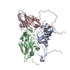
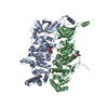
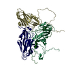
| Entry | Database: PDB / ID: 4kf7 | ||||||
|---|---|---|---|---|---|---|---|
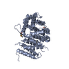
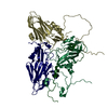
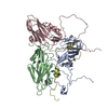
| Title | Nup188(aa1-1160) from Myceliophthora thermophila | ||||||
 Components Components | Nup188 Nucleoporin 188 Nucleoporin 188 | ||||||
 Keywords Keywords | STRUCTURAL PROTEIN / Nucleoporin | ||||||
| Function / homology |  Function and homology information Function and homology informationstructural constituent of nuclear pore / nuclear pore / protein transportSimilarity search - Function | ||||||
| Biological species |  Myceliophthora thermophila (fungus) Myceliophthora thermophila (fungus) | ||||||
| Method | X-RAY DIFFRACTION / SYNCHROTRON / SAD / Resolution: 2.65 Å | ||||||
 Authors Authors | Schwartz, T.U. / Andersen, K.R. | ||||||
 Citation Citation | Journal: Elife / Year: 2013 Title: Scaffold nucleoporins Nup188 and Nup192 share structural and functional properties with nuclear transport receptors. Authors: Andersen, K.R. / Onischenko, E. / Tang, J.H. / Kumar, P. / Chen, J.Z. / Ulrich, A. / Liphardt, J.T. / Weis, K. / Schwartz, T.U. | ||||||
| History |
|
- Structure visualization
| Structure viewer | Molecule: MolmilJmol/JSmol |
|---|
- Downloads & links
-Download
| PDBx/mmCIF format | 4kf7.cif.gz | 442.7 KB | Display | PDBx/mmCIF format |
|---|---|---|---|---|
| PDB format | pdb4kf7.ent.gz | 364.7 KB | Display | PDB format |
| PDBx/mmJSON format | 4kf7.json.gz | Tree view | PDBx/mmJSON format | |
| Others |  Other downloads Other downloads |
-Validation report
| Arichive directory | https://data.pdbj.org/pub/pdb/validation_reports/kf/4kf7ftp://data.pdbj.org/pub/pdb/validation_reports/kf/4kf7 | HTTPS FTP |
|---|
-Related structure data


-Links
 PDBj
PDBj- Assembly
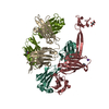
| Deposited unit | 
| ||||||||
|---|---|---|---|---|---|---|---|---|---|
| 1 |
| ||||||||
| Unit cell |
|
-Components
| #1: Protein | Nucleoporin 188 Mass: 128246.492 Da / Num. of mol.: 1 / Fragment: N-terminal domain (UNP residues 1-1160) Source method: isolated from a genetically manipulated source Source: (gene. exp.) Myceliophthora thermophila (fungus) / Gene: MYCTH_2303581 / Production host:  Escherichia coli (E. coli) / References: UniProt: G2QD05 Escherichia coli (E. coli) / References: UniProt: G2QD05 |
|---|---|
| #2: Water | ChemComp-HOH / Water Mass: 18.015 Da / Num. of mol.: 128 / Source method: isolated from a natural source / Formula: H2O Mass: 18.015 Da / Num. of mol.: 128 / Source method: isolated from a natural source / Formula: H2O |
-Experimental details
-Experiment
| Experiment | Method: X-RAY DIFFRACTION / Number of used crystals: 1 |
|---|
- Sample preparation
| Crystal | Density Matthews: 2.83 Å3/Da / Density % sol: 56.49 % |
|---|---|
| Crystal grow | Temperature: 290 K / Method: vapor diffusion, hanging drop / pH: 6.5 Details: 0.1 M MES, 4.5-7.0% w/v PEG4000, 150 mM ammonium sulfate, 1 mM DTT, 1.0-2.5% tert-butanol, pH 6.5, VAPOR DIFFUSION, HANGING DROP, temperature 290K |
-Data collection
| Diffraction | Mean temperature: 100 K |
|---|---|
| Diffraction source | Source: SYNCHROTRON / Site: APS  / Beamline: 24-ID-C / Wavelength: 0.9792 Å / Beamline: 24-ID-C / Wavelength: 0.9792 Å |
| Detector | Type: PSI PILATUS 6M / Detector: PIXEL / Date: Mar 3, 2012 |
| Radiation | Monochromator: double crystal Si(111) / Protocol: SINGLE WAVELENGTH / Monochromatic (M) / Laue (L): M / Scattering type: x-ray |
| Radiation wavelength | Wavelength: 0.9792 Å / Relative weight: 1 |
| Reflection | Resolution: 2.65→66.8 Å / Num. all: 41673 / Num. obs: 41673 / % possible obs: 100 % / Observed criterion σ(F): 1.8 / Observed criterion σ(I): 1.8 / Redundancy: 4.1 % / Rmerge(I) obs: 0.031 / Net I/σ(I): 31.3 |
| Reflection shell | Resolution: 2.65→2.72 Å / Redundancy: 3.8 % / Rmerge(I) obs: 0.772 / Mean I/σ(I) obs: 1.8 / % possible all: 99.9 |
- Processing
| Software |
| ||||||||||||||||||||||||||||||||||||||||||||||||||||||||||||||||||||||||||||||||||||||||||||||||||||||||||||||||||||||||||||||||||||||||||||||||||||||
|---|---|---|---|---|---|---|---|---|---|---|---|---|---|---|---|---|---|---|---|---|---|---|---|---|---|---|---|---|---|---|---|---|---|---|---|---|---|---|---|---|---|---|---|---|---|---|---|---|---|---|---|---|---|---|---|---|---|---|---|---|---|---|---|---|---|---|---|---|---|---|---|---|---|---|---|---|---|---|---|---|---|---|---|---|---|---|---|---|---|---|---|---|---|---|---|---|---|---|---|---|---|---|---|---|---|---|---|---|---|---|---|---|---|---|---|---|---|---|---|---|---|---|---|---|---|---|---|---|---|---|---|---|---|---|---|---|---|---|---|---|---|---|---|---|---|---|---|---|---|---|---|
| Refinement | Method to determine structure: SAD / Resolution: 2.65→66.759 Å / SU ML: 0.34 / σ(F): 1.8 / Phase error: 24.77 / Stereochemistry target values: ML
| ||||||||||||||||||||||||||||||||||||||||||||||||||||||||||||||||||||||||||||||||||||||||||||||||||||||||||||||||||||||||||||||||||||||||||||||||||||||
| Solvent computation | Shrinkage radii: 0.9 Å / VDW probe radii: 1.11 Å / Solvent model: FLAT BULK SOLVENT MODEL | ||||||||||||||||||||||||||||||||||||||||||||||||||||||||||||||||||||||||||||||||||||||||||||||||||||||||||||||||||||||||||||||||||||||||||||||||||||||
| Refinement step | Cycle: LAST / Resolution: 2.65→66.759 Å
| ||||||||||||||||||||||||||||||||||||||||||||||||||||||||||||||||||||||||||||||||||||||||||||||||||||||||||||||||||||||||||||||||||||||||||||||||||||||
| Refine LS restraints |
| ||||||||||||||||||||||||||||||||||||||||||||||||||||||||||||||||||||||||||||||||||||||||||||||||||||||||||||||||||||||||||||||||||||||||||||||||||||||
| LS refinement shell |
| ||||||||||||||||||||||||||||||||||||||||||||||||||||||||||||||||||||||||||||||||||||||||||||||||||||||||||||||||||||||||||||||||||||||||||||||||||||||
| Refinement TLS params. | Method: refined / Refine-ID: X-RAY DIFFRACTION
| ||||||||||||||||||||||||||||||||||||||||||||||||||||||||||||||||||||||||||||||||||||||||||||||||||||||||||||||||||||||||||||||||||||||||||||||||||||||
| Refinement TLS group |
|
