 Movie
Movie Controller


+ Open data

- Basic information
| Entry | Database: PDB / ID: 4k25 | ||||||
|---|---|---|---|---|---|---|---|
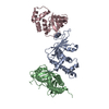
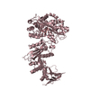
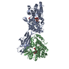
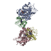
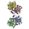
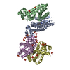
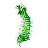
| Title | Crystal Structure of yeast Qri7 homodimer | ||||||
 Components Components | Probable tRNA threonylcarbamoyladenosine biosynthesis protein QRI7, mitochondrial | ||||||
 Keywords Keywords |  BIOSYNTHETIC PROTEIN / ASKHA-fold / N6-threonylcarbamoylation enzyme / RNA and metabolite binding / mitochondria BIOSYNTHETIC PROTEIN / ASKHA-fold / N6-threonylcarbamoylation enzyme / RNA and metabolite binding / mitochondria | ||||||
| Function / homology |  Function and homology information Function and homology informationmitochondrial tRNA threonylcarbamoyladenosine modification / N6-L-threonylcarbamoyladenine synthase / N(6)-L-threonylcarbamoyladenine synthase activity / tRNA threonylcarbamoyladenosine modification / mitochondrion / metal ion bindingSimilarity search - Function | ||||||
| Biological species |  Saccharomyces cerevisiae (brewer's yeast) Saccharomyces cerevisiae (brewer's yeast) | ||||||
| Method | X-RAY DIFFRACTION / SYNCHROTRON / MOLECULAR REPLACEMENT / Resolution: 2.88 Å | ||||||
 Authors Authors | Neculai, D. / Wan, L. / Mao, D.Y. / Sicheri, F. | ||||||
 Citation Citation | Journal: Nucleic Acids Res. / Year: 2013 Title: Reconstitution and characterization of eukaryotic N6-threonylcarbamoylation of tRNA using a minimal enzyme system. Authors: Wan, L.C. / Mao, D.Y. / Neculai, D. / Strecker, J. / Chiovitti, D. / Kurinov, I. / Poda, G. / Thevakumaran, N. / Yuan, F. / Szilard, R.K. / Lissina, E. / Nislow, C. / Caudy, A.A. / Durocher, D. / Sicheri, F. | ||||||
| History |
|
- Structure visualization
| Structure viewer | Molecule: MolmilJmol/JSmol |
|---|
- Downloads & links
-Download
| PDBx/mmCIF format | 4k25.cif.gz | 148.7 KB | Display | PDBx/mmCIF format |
|---|---|---|---|---|
| PDB format | pdb4k25.ent.gz | 123.5 KB | Display | PDB format |
| PDBx/mmJSON format | 4k25.json.gz | Tree view | PDBx/mmJSON format | |
| Others |  Other downloads Other downloads |
-Validation report
| Arichive directory | https://data.pdbj.org/pub/pdb/validation_reports/k2/4k25ftp://data.pdbj.org/pub/pdb/validation_reports/k2/4k25 | HTTPS FTP |
|---|
-Related structure data
| Similar structure data |
|---|


-Links
 PDBj
PDBj- Assembly
| Deposited unit | 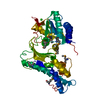
| ||||||||
|---|---|---|---|---|---|---|---|---|---|
| 1 | 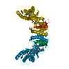
| ||||||||
| Unit cell |
| ||||||||
| Details | The second part of the biological assembly is generated by the two-fold axis: -X, -Y, Z + (1 0 0) |
-Components
| #1: Protein | Mass: 42989.711 Da / Num. of mol.: 1 Source method: isolated from a genetically manipulated source Source: (gene. exp.) Saccharomyces cerevisiae (brewer's yeast)Strain: ATCC 204508 / S288c / Gene: QRI7, YDL104C, D2366 / Production host:  Escherichia coli (E. coli) / References: UniProt: P43122 Escherichia coli (E. coli) / References: UniProt: P43122 |
|---|---|
| #2: Chemical | ChemComp-ZN /   Mass: 65.409 Da / Num. of mol.: 1 / Source method: obtained synthetically / Formula: Zn Mass: 65.409 Da / Num. of mol.: 1 / Source method: obtained synthetically / Formula: Zn |
| #3: Chemical | ChemComp-CA /   Mass: 40.078 Da / Num. of mol.: 1 / Source method: obtained synthetically / Formula: Ca Mass: 40.078 Da / Num. of mol.: 1 / Source method: obtained synthetically / Formula: Ca |
-Experimental details
-Experiment
| Experiment | Method: X-RAY DIFFRACTION / Number of used crystals: 1 |
|---|
- Sample preparation
| Crystal | Density Matthews: 2.36 Å3/Da / Density % sol: 47.77 % |
|---|---|
| Crystal grow | Temperature: 277.15 K / Method: vapor diffusion, hanging drop / pH: 7.5 Details: 0.1M Tris, 8% PEG8000, 0.2M magnesium chloride, pH 7.5, VAPOR DIFFUSION, HANGING DROP, temperature 277.15K |
-Data collection
| Diffraction | Mean temperature: 100 K |
|---|---|
| Diffraction source | Source: SYNCHROTRON / Site: APS  / Beamline: 24-ID-E / Wavelength: 0.97933 Å / Beamline: 24-ID-E / Wavelength: 0.97933 Å |
| Detector | Type: ADSC QUANTUM 315 / Detector: CCD / Date: Jul 20, 2011 |
| Radiation | Monochromator: Si (220), Si (311) / Protocol: SINGLE WAVELENGTH / Monochromatic (M) / Laue (L): M / Scattering type: x-ray |
| Radiation wavelength | Wavelength: 0.97933 Å / Relative weight: 1 |
| Reflection | Resolution: 2.88→20.58 Å / Num. all: 17164 / Num. obs: 9434 / % possible obs: 55 % / Observed criterion σ(F): 2.54 / Observed criterion σ(I): 2.54 / Biso Wilson estimate: 77.86 Å2 |
| Reflection shell | Resolution: 2.88→2.98 Å / % possible all: 99.6 |
- Processing
| Software |
| ||||||||||||||||||||||||||||||||||||||||||||||||||||||||||||||||||||||||
|---|---|---|---|---|---|---|---|---|---|---|---|---|---|---|---|---|---|---|---|---|---|---|---|---|---|---|---|---|---|---|---|---|---|---|---|---|---|---|---|---|---|---|---|---|---|---|---|---|---|---|---|---|---|---|---|---|---|---|---|---|---|---|---|---|---|---|---|---|---|---|---|---|---|
| Refinement | Method to determine structure: MOLECULAR REPLACEMENT / Resolution: 2.88→20.58 Å / Cor.coef. Fo:Fc: 0.9059 / Cor.coef. Fo:Fc free: 0.8745 / Cross valid method: THROUGHOUT / σ(F): 3.3 / Stereochemistry target values: Engh & Huber
| ||||||||||||||||||||||||||||||||||||||||||||||||||||||||||||||||||||||||
| Displacement parameters | Biso mean: 81.54 Å2
| ||||||||||||||||||||||||||||||||||||||||||||||||||||||||||||||||||||||||
| Refine analyze | Luzzati coordinate error obs: 0.625 Å | ||||||||||||||||||||||||||||||||||||||||||||||||||||||||||||||||||||||||
| Refinement step | Cycle: LAST / Resolution: 2.88→20.58 Å
| ||||||||||||||||||||||||||||||||||||||||||||||||||||||||||||||||||||||||
| Refine LS restraints |
| ||||||||||||||||||||||||||||||||||||||||||||||||||||||||||||||||||||||||
| LS refinement shell | Resolution: 2.9→3.24 Å / Total num. of bins used: 5
| ||||||||||||||||||||||||||||||||||||||||||||||||||||||||||||||||||||||||
| Refinement TLS params. | Method: refined / Origin x: 46.0749 Å / Origin y: 17.508 Å / Origin z: 24.6981 Å
| ||||||||||||||||||||||||||||||||||||||||||||||||||||||||||||||||||||||||
| Refinement TLS group | Selection details: { A|33 - A|602 } |
