 Movie
Movie Controller


+ Open data

- Basic information
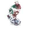
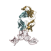
| Entry | Database: PDB / ID: 4k17 | |||||||||
|---|---|---|---|---|---|---|---|---|---|---|
| Title | Crystal Structure of mouse CARMIL residues 1-668 | |||||||||
 Components Components | Leucine-rich repeat-containing protein 16A | |||||||||
 Keywords Keywords | LIPID BINDING PROTEIN /  PH domain / LRR domain / lipid binding / protein-protein interaction / phosphatidylserine / phosphatidylinositol / phosphatidylinositol-5-phosphate / plasma membrane PH domain / LRR domain / lipid binding / protein-protein interaction / phosphatidylserine / phosphatidylinositol / phosphatidylinositol-5-phosphate / plasma membrane | |||||||||
| Function / homology |  Function and homology information Function and homology informationbarbed-end actin filament uncapping / positive regulation of lamellipodium organization / negative regulation of barbed-end actin filament capping / macropinosome / actin filament network formation / macropinocytosis / urate metabolic process / regulation of Arp2/3 complex-mediated actin nucleation / Factors involved in megakaryocyte development and platelet production / ruffle organization ...barbed-end actin filament uncapping / positive regulation of lamellipodium organization / negative regulation of barbed-end actin filament capping / macropinosome / actin filament network formation / macropinocytosis / urate metabolic process / regulation of Arp2/3 complex-mediated actin nucleation / Factors involved in megakaryocyte development and platelet production / ruffle organization / intermediate filament cytoskeleton / lamellipodium assembly / establishment or maintenance of cell polarity / positive regulation of actin filament polymerization / cell leading edge / filamentous actin / positive regulation of substrate adhesion-dependent cell spreading / positive regulation of stress fiber assembly / cell migration / lamellipodium / positive regulation of cell migration / nuclear speck / protein-containing complex binding / nucleoplasm / nucleus / plasma membrane / cytosol / cytoplasmSimilarity search - Function | |||||||||
| Biological species |  Mus musculus (house mouse) Mus musculus (house mouse) | |||||||||
| Method | X-RAY DIFFRACTION / SYNCHROTRON / SAD / Resolution: 2.895 Å | |||||||||
 Authors Authors | Zwolak, A. / Dominguez, R. | |||||||||
 Citation Citation | Journal: Nat Commun / Year: 2013 Title: CARMIL leading edge localization depends on a non-canonical PH domain and dimerization. Authors: Zwolak, A. / Yang, C. / Feeser, E.A. / Michael Ostap, E. / Svitkina, T. / Dominguez, R. | |||||||||
| History |
|
- Structure visualization
| Structure viewer | Molecule: MolmilJmol/JSmol |
|---|
- Downloads & links
-Download
| PDBx/mmCIF format | 4k17.cif.gz | 503.6 KB | Display | PDBx/mmCIF format |
|---|---|---|---|---|
| PDB format | pdb4k17.ent.gz | 418.8 KB | Display | PDB format |
| PDBx/mmJSON format | 4k17.json.gz | Tree view | PDBx/mmJSON format | |
| Others |  Other downloads Other downloads |
-Validation report
| Arichive directory | https://data.pdbj.org/pub/pdb/validation_reports/k1/4k17ftp://data.pdbj.org/pub/pdb/validation_reports/k1/4k17 | HTTPS FTP |
|---|
-Related structure data
| Similar structure data |
|---|


-Links
 PDBj
PDBj


- Assembly
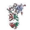
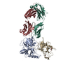
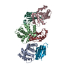
| Deposited unit | 
| ||||||||
|---|---|---|---|---|---|---|---|---|---|
| 1 | 
| ||||||||
| 2 | 
| ||||||||
| 3 | 
| ||||||||
| 4 | 
| ||||||||
| Unit cell |
|
-Components
| #1: Protein | Mass: 74778.203 Da / Num. of mol.: 4 / Fragment: PH and LRR domains (UNP residues 1-668) Source method: isolated from a genetically manipulated source Source: (gene. exp.) Mus musculus (house mouse) / Gene: Carmil, Lrrc16, Lrrc16a / Plasmid: pRSFDuet-1 / Production host:  Escherichia coli (E. coli) / Strain (production host): BL21 RIPL / References: UniProt: Q6EDY6 Escherichia coli (E. coli) / Strain (production host): BL21 RIPL / References: UniProt: Q6EDY6#2: Chemical | ChemComp-OHB / | Salicylamide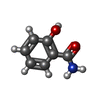  Mass: 137.136 Da / Num. of mol.: 1 / Source method: obtained synthetically / Formula: C7H7NO2 Mass: 137.136 Da / Num. of mol.: 1 / Source method: obtained synthetically / Formula: C7H7NO2#3: Chemical | ChemComp-CL / | Chloride  Mass: 35.453 Da / Num. of mol.: 1 / Source method: obtained synthetically / Formula: Cl Mass: 35.453 Da / Num. of mol.: 1 / Source method: obtained synthetically / Formula: Cl#4: Chemical | ChemComp-ABU / | Γ-Aminobutyric acid  Mass: 103.120 Da / Num. of mol.: 1 / Source method: obtained synthetically / Formula: C4H9NO2 / Comment: neurotransmitter, inhibitor*YM Mass: 103.120 Da / Num. of mol.: 1 / Source method: obtained synthetically / Formula: C4H9NO2 / Comment: neurotransmitter, inhibitor*YM |
|---|
-Experimental details
-Experiment
| Experiment | Method: X-RAY DIFFRACTION / Number of used crystals: 1 |
|---|
- Sample preparation
| Crystal | Density Matthews: 2.56 Å3/Da / Density % sol: 51.92 % |
|---|---|
| Crystal grow | Temperature: 293 K / Method: vapor diffusion, hanging drop / pH: 7.7 Details: 16 % PEG3350, 130 mM lithium sulfate, 0.25 % w/v salicylamide, pH 7.7, VAPOR DIFFUSION, HANGING DROP, temperature 293K |
-Data collection
| Diffraction | Mean temperature: 100 K |
|---|---|
| Diffraction source | Source: SYNCHROTRON / Site: NSLS  / Beamline: X6A / Wavelength: 0.9784 Å / Beamline: X6A / Wavelength: 0.9784 Å |
| Detector | Type: ADSC QUANTUM 270 / Detector: CCD / Date: Aug 4, 2012 / Details: toroidal focusing mirror |
| Radiation | Monochromator: Si(111) channel cut / Protocol: SINGLE WAVELENGTH / Monochromatic (M) / Laue (L): M / Scattering type: x-ray |
| Radiation wavelength | Wavelength: 0.9784 Å / Relative weight: 1 |
| Reflection | Resolution: 2.895→50 Å / Num. all: 65054 / Num. obs: 63752 / % possible obs: 98 % / Observed criterion σ(F): 2 / Observed criterion σ(I): 2 / Redundancy: 8 % / Rmerge(I) obs: 0.124 / Net I/σ(I): 13.8 |
| Reflection shell | Resolution: 2.895→3 Å / Redundancy: 7.4 % / Rmerge(I) obs: 0.825 / Mean I/σ(I) obs: 1.8 / % possible all: 84.4 |
- Processing
| Software |
| ||||||||||||||||||||
|---|---|---|---|---|---|---|---|---|---|---|---|---|---|---|---|---|---|---|---|---|---|
| Refinement | Method to determine structure: SAD / Resolution: 2.895→49.759 Å / Cor.coef. Fo:Fc: 0.9048 / σ(F): 2 / Stereochemistry target values: Engh & Huber
| ||||||||||||||||||||
| Refinement step | Cycle: LAST / Resolution: 2.895→49.759 Å
| ||||||||||||||||||||
| Refine LS restraints |
|
