 Movie
Movie Controller

[English] 日本語
 Yorodumi
- PDB-4jz5: High-resolution structure of catalytic domain of endolysin ply40 ... -
+ Open data

- Basic information
| Entry | Database: PDB / ID: 4jz5 | ||||||
|---|---|---|---|---|---|---|---|
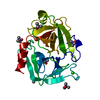
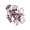
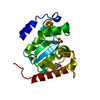
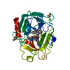
| Title | High-resolution structure of catalytic domain of endolysin ply40 from bacteriophage P40 of Listeria monocytogenes | ||||||
 Components Components | Gp26 | ||||||
 Keywords Keywords |  HYDROLASE / Endolysin / Glycosyl hydrolase family 25 / Peptidoglycan / cell-wall HYDROLASE / Endolysin / Glycosyl hydrolase family 25 / Peptidoglycan / cell-wall | ||||||
| Function / homology |  Function and homology information Function and homology informationpeptidoglycan catabolic process / cell wall macromolecule catabolic process / lysozyme activitySimilarity search - Function | ||||||
| Biological species |  Listeria phage P40 (virus) Listeria phage P40 (virus) | ||||||
| Method | X-RAY DIFFRACTION / SYNCHROTRON / MOLECULAR REPLACEMENT / Resolution: 1.1 Å | ||||||
 Authors Authors | Romero-Fernandez, P. / Bartual, S.G. / Carrasco-lopez, C. / Hermoso, J.A. | ||||||
 Citation Citation | Journal: To be Published Title: Crystal structure of catalytic domain of endolysin Ply40 Authors: Romero-Fernandez, P. / Bartual, S.G. / Carrasco-lopez, C. / Loessner, M. / Hermoso, J.A. | ||||||
| History |
|
- Structure visualization
| Structure viewer | Molecule: MolmilJmol/JSmol |
|---|
- Downloads & links
-Download
| PDBx/mmCIF format | 4jz5.cif.gz | 138.2 KB | Display | PDBx/mmCIF format |
|---|---|---|---|---|
| PDB format | pdb4jz5.ent.gz | 108.4 KB | Display | PDB format |
| PDBx/mmJSON format | 4jz5.json.gz | Tree view | PDBx/mmJSON format | |
| Others |  Other downloads Other downloads |
-Validation report
| Arichive directory | https://data.pdbj.org/pub/pdb/validation_reports/jz/4jz5ftp://data.pdbj.org/pub/pdb/validation_reports/jz/4jz5 | HTTPS FTP |
|---|
-Related structure data
| Related structure data | 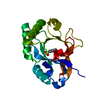 3hmcS S: Starting model for refinement |
|---|---|
| Similar structure data |


-Links
 PDBj
PDBj
- Assembly
| Deposited unit | 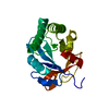
| ||||||||
|---|---|---|---|---|---|---|---|---|---|
| 1 |
| ||||||||
| Unit cell |
|
-Components
| #1: Protein | Mass: 25078.514 Da / Num. of mol.: 1 / Fragment: catalytic domain, UNP residues 1-202 Source method: isolated from a genetically manipulated source Source: (gene. exp.) Listeria phage P40 (virus) / Gene: gp26 / Production host:  Escherichia coli (E. coli) / References: UniProt: B6D7J9 Escherichia coli (E. coli) / References: UniProt: B6D7J9 |
|---|---|
| #2: Water | ChemComp-HOH / Water Mass: 18.015 Da / Num. of mol.: 345 / Source method: isolated from a natural source / Formula: H2O Mass: 18.015 Da / Num. of mol.: 345 / Source method: isolated from a natural source / Formula: H2O |
-Experimental details
-Experiment
| Experiment | Method: X-RAY DIFFRACTION / Number of used crystals: 1 |
|---|
- Sample preparation
| Crystal | Density Matthews: 2.03 Å3/Da / Density % sol: 39.54 % |
|---|---|
| Crystal grow | pH: 7 / Details: 0.2M Sodium Malonate, 20%(w/v) PEG, pH 7.0 |
-Data collection
| Diffraction | Mean temperature: 100 K |
|---|---|
| Diffraction source | Source: SYNCHROTRON / Site: ESRF  / Beamline: ID14-1 / Wavelength: 0.9334 Å / Beamline: ID14-1 / Wavelength: 0.9334 Å |
| Detector | Type: ADSC QUANTUM 315r / Detector: CCD / Date: Nov 8, 2011 |
| Radiation | Monochromator: NA / Protocol: SINGLE WAVELENGTH / Monochromatic (M) / Laue (L): M / Scattering type: x-ray |
| Radiation wavelength | Wavelength: 0.9334 Å / Relative weight: 1 |
| Reflection | Resolution: 1.1→36.661 Å / Num. all: 78926 / Num. obs: 78926 / % possible obs: 98.2 % / Observed criterion σ(F): 0 / Observed criterion σ(I): 2 |
| Reflection shell | Resolution: 1.1→1.12 Å / % possible all: 80 |
- Processing
| Software |
| |||||||||||||||||||||||||||||||||||||||||||||||||||||||||||||||||||||||||||||||||||||||||||||||||||||||||||||||||||||||||||||||||||||||||||||||||||||||||||||||||||||||||||||||||||||||||||||||||||||||||||||||||||||||||||||||||||||||||||||||||||||||||||||||||||||||||||||||||||
|---|---|---|---|---|---|---|---|---|---|---|---|---|---|---|---|---|---|---|---|---|---|---|---|---|---|---|---|---|---|---|---|---|---|---|---|---|---|---|---|---|---|---|---|---|---|---|---|---|---|---|---|---|---|---|---|---|---|---|---|---|---|---|---|---|---|---|---|---|---|---|---|---|---|---|---|---|---|---|---|---|---|---|---|---|---|---|---|---|---|---|---|---|---|---|---|---|---|---|---|---|---|---|---|---|---|---|---|---|---|---|---|---|---|---|---|---|---|---|---|---|---|---|---|---|---|---|---|---|---|---|---|---|---|---|---|---|---|---|---|---|---|---|---|---|---|---|---|---|---|---|---|---|---|---|---|---|---|---|---|---|---|---|---|---|---|---|---|---|---|---|---|---|---|---|---|---|---|---|---|---|---|---|---|---|---|---|---|---|---|---|---|---|---|---|---|---|---|---|---|---|---|---|---|---|---|---|---|---|---|---|---|---|---|---|---|---|---|---|---|---|---|---|---|---|---|---|---|---|---|---|---|---|---|---|---|---|---|---|---|---|---|---|---|---|---|---|---|---|---|---|---|---|---|---|---|---|---|---|---|---|---|---|---|---|---|---|---|---|---|---|---|---|---|---|---|---|
| Refinement | Method to determine structure: MOLECULAR REPLACEMENT Starting model: 3HMC Resolution: 1.1→36.661 Å / Occupancy max: 1 / Occupancy min: 0.44 / SU ML: 0.07 / σ(F): 1.98 / Phase error: 12.89 / Stereochemistry target values: ML
| |||||||||||||||||||||||||||||||||||||||||||||||||||||||||||||||||||||||||||||||||||||||||||||||||||||||||||||||||||||||||||||||||||||||||||||||||||||||||||||||||||||||||||||||||||||||||||||||||||||||||||||||||||||||||||||||||||||||||||||||||||||||||||||||||||||||||||||||||||
| Solvent computation | Shrinkage radii: 0.9 Å / VDW probe radii: 1.11 Å / Solvent model: FLAT BULK SOLVENT MODEL | |||||||||||||||||||||||||||||||||||||||||||||||||||||||||||||||||||||||||||||||||||||||||||||||||||||||||||||||||||||||||||||||||||||||||||||||||||||||||||||||||||||||||||||||||||||||||||||||||||||||||||||||||||||||||||||||||||||||||||||||||||||||||||||||||||||||||||||||||||
| Displacement parameters | Biso max: 48.39 Å2 / Biso mean: 11.511 Å2 / Biso min: 3.53 Å2 | |||||||||||||||||||||||||||||||||||||||||||||||||||||||||||||||||||||||||||||||||||||||||||||||||||||||||||||||||||||||||||||||||||||||||||||||||||||||||||||||||||||||||||||||||||||||||||||||||||||||||||||||||||||||||||||||||||||||||||||||||||||||||||||||||||||||||||||||||||
| Refinement step | Cycle: LAST / Resolution: 1.1→36.661 Å
| |||||||||||||||||||||||||||||||||||||||||||||||||||||||||||||||||||||||||||||||||||||||||||||||||||||||||||||||||||||||||||||||||||||||||||||||||||||||||||||||||||||||||||||||||||||||||||||||||||||||||||||||||||||||||||||||||||||||||||||||||||||||||||||||||||||||||||||||||||
| Refine LS restraints |
| |||||||||||||||||||||||||||||||||||||||||||||||||||||||||||||||||||||||||||||||||||||||||||||||||||||||||||||||||||||||||||||||||||||||||||||||||||||||||||||||||||||||||||||||||||||||||||||||||||||||||||||||||||||||||||||||||||||||||||||||||||||||||||||||||||||||||||||||||||
| LS refinement shell | Refine-ID: X-RAY DIFFRACTION / Total num. of bins used: 28
| |||||||||||||||||||||||||||||||||||||||||||||||||||||||||||||||||||||||||||||||||||||||||||||||||||||||||||||||||||||||||||||||||||||||||||||||||||||||||||||||||||||||||||||||||||||||||||||||||||||||||||||||||||||||||||||||||||||||||||||||||||||||||||||||||||||||||||||||||||
| Refinement TLS params. | Method: refined / Refine-ID: X-RAY DIFFRACTION
| |||||||||||||||||||||||||||||||||||||||||||||||||||||||||||||||||||||||||||||||||||||||||||||||||||||||||||||||||||||||||||||||||||||||||||||||||||||||||||||||||||||||||||||||||||||||||||||||||||||||||||||||||||||||||||||||||||||||||||||||||||||||||||||||||||||||||||||||||||
| Refinement TLS group |
|
