 Movie
Movie Controller


+ Open data

- Basic information
| Entry | Database: PDB / ID: 4jvc | ||||||
|---|---|---|---|---|---|---|---|
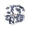
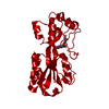
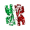
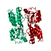
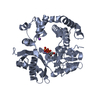
| Title | Crystal structure of PqsR co-inducer binding domain | ||||||
 Components Components | Transcriptional regulator MvfR | ||||||
 Keywords Keywords |  TRANSCRIPTION REGULATOR / transcription regulation / co-inducer binding / DNA binding TRANSCRIPTION REGULATOR / transcription regulation / co-inducer binding / DNA binding | ||||||
| Function / homology |  Function and homology information Function and homology information | ||||||
| Biological species |   Pseudomonas aeruginosa (bacteria) Pseudomonas aeruginosa (bacteria) | ||||||
| Method | X-RAY DIFFRACTION / SYNCHROTRON / MIRAS / Resolution: 2.5 Å | ||||||
 Authors Authors | Ilangovan, A. / Emsley, J. / Williams, P. | ||||||
 Citation Citation | Journal: Plos Pathog. / Year: 2013 Title: Structural basis for native agonist and synthetic inhibitor recognition by the Pseudomonas aeruginosa quorum sensing regulator PqsR (MvfR). Authors: Ilangovan, A. / Fletcher, M. / Rampioni, G. / Pustelny, C. / Rumbaugh, K. / Heeb, S. / Camara, M. / Truman, A. / Chhabra, S.R. / Emsley, J. / Williams, P. | ||||||
| History |
|
- Structure visualization
| Structure viewer | Molecule: MolmilJmol/JSmol |
|---|
- Downloads & links
-Download
| PDBx/mmCIF format | 4jvc.cif.gz | 53.8 KB | Display | PDBx/mmCIF format |
|---|---|---|---|---|
| PDB format | pdb4jvc.ent.gz | 38.9 KB | Display | PDB format |
| PDBx/mmJSON format | 4jvc.json.gz | Tree view | PDBx/mmJSON format | |
| Others |  Other downloads Other downloads |
-Validation report
| Arichive directory | https://data.pdbj.org/pub/pdb/validation_reports/jv/4jvcftp://data.pdbj.org/pub/pdb/validation_reports/jv/4jvc | HTTPS FTP |
|---|
-Related structure data


-Links
 PDBj
PDBj- Assembly
| Deposited unit | 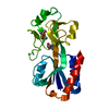
| ||||||||
|---|---|---|---|---|---|---|---|---|---|
| 1 | 
| ||||||||
| Unit cell |
|
-Components
| #1: Protein | Mass: 24288.475 Da / Num. of mol.: 1 / Fragment: co-inducer binding domain, UNP RESIDUES 94-309 Source method: isolated from a genetically manipulated source Source: (gene. exp.) Pseudomonas aeruginosa (bacteria) / Strain: UCBPP-PA14 / Gene: mvfR, PA14_51340, PqsR / Plasmid: pET28a / Production host: Escherichia coli (E. coli) / Strain (production host): BL21 DE3 / References: UniProt: Q02IG8, UniProt: A0A0H2Z7A6*PLUS | ||
|---|---|---|---|
| #2: Chemical | 2-Methyl-2,4-pentanediol  Mass: 118.174 Da / Num. of mol.: 2 / Source method: obtained synthetically / Formula: C6H14O2 / Comment: precipitant*YM Mass: 118.174 Da / Num. of mol.: 2 / Source method: obtained synthetically / Formula: C6H14O2 / Comment: precipitant*YM#3: Water | ChemComp-HOH / | Water Mass: 18.015 Da / Num. of mol.: 3 / Source method: isolated from a natural source / Formula: H2O Mass: 18.015 Da / Num. of mol.: 3 / Source method: isolated from a natural source / Formula: H2O |
-Experimental details
-Experiment
| Experiment | Method: X-RAY DIFFRACTION / Number of used crystals: 1 |
|---|
- Sample preparation
| Crystal | Density Matthews: 4.93 Å3/Da / Density % sol: 75.06 % |
|---|---|
| Crystal grow | Temperature: 292 K / Method: vapor diffusion, sitting drop / pH: 6.5 Details: 0.1M Ammonium acetate, 0.1M Tri sodium citrate pH 6.5, 3% MPD, VAPOR DIFFUSION, SITTING DROP, temperature 292K |
-Data collection
| Diffraction | Mean temperature: 100 K | |||||||||||||||||||||||||||||||||||||||
|---|---|---|---|---|---|---|---|---|---|---|---|---|---|---|---|---|---|---|---|---|---|---|---|---|---|---|---|---|---|---|---|---|---|---|---|---|---|---|---|---|
| Diffraction source | Source: SYNCHROTRON / Site: Diamond  / Beamline: I04 / Wavelength: 0.977 Å / Beamline: I04 / Wavelength: 0.977 Å | |||||||||||||||||||||||||||||||||||||||
| Detector | Type: ADSC QUANTUM 315r / Detector: CCD | |||||||||||||||||||||||||||||||||||||||
| Radiation | Monochromator: GRAPHITE / Monochromatic (M) / Laue (L): M / Scattering type: x-ray | |||||||||||||||||||||||||||||||||||||||
| Radiation wavelength | Wavelength: 0.977 Å / Relative weight: 1 | |||||||||||||||||||||||||||||||||||||||
| Reflection | Resolution: 2.5→30 Å / Num. all: 19299 / Num. obs: 17339 / % possible obs: 99.2 % / Observed criterion σ(F): 1 / Observed criterion σ(I): 1 / Redundancy: 7.9 % / Biso Wilson estimate: 71.9 Å2 / Rmerge(I) obs: 0.74 / Rsym value: 0.85 / Net I/σ(I): 2 | |||||||||||||||||||||||||||||||||||||||
| Reflection shell |
|
- Processing
| Software |
| ||||||||||||||||||||||||||||||||||||||||||||||||||||||||||||
|---|---|---|---|---|---|---|---|---|---|---|---|---|---|---|---|---|---|---|---|---|---|---|---|---|---|---|---|---|---|---|---|---|---|---|---|---|---|---|---|---|---|---|---|---|---|---|---|---|---|---|---|---|---|---|---|---|---|---|---|---|---|
| Refinement | Method to determine structure: MIRAS / Resolution: 2.5→28.98 Å / Cor.coef. Fo:Fc: 0.957 / Cor.coef. Fo:Fc free: 0.94 / SU B: 11.112 / SU ML: 0.225 / Cross valid method: THROUGHOUT / ESU R: 0.228 / ESU R Free: 0.214 / Stereochemistry target values: MAXIMUM LIKELIHOOD / Details: HYDROGENS HAVE BEEN ADDED IN THE RIDING POSITIONS
| ||||||||||||||||||||||||||||||||||||||||||||||||||||||||||||
| Solvent computation | Ion probe radii: 0.8 Å / Shrinkage radii: 0.8 Å / VDW probe radii: 1.2 Å / Solvent model: MASK | ||||||||||||||||||||||||||||||||||||||||||||||||||||||||||||
| Displacement parameters | Biso mean: 99.889 Å2
| ||||||||||||||||||||||||||||||||||||||||||||||||||||||||||||
| Refinement step | Cycle: LAST / Resolution: 2.5→28.98 Å
| ||||||||||||||||||||||||||||||||||||||||||||||||||||||||||||
| Refine LS restraints |
| ||||||||||||||||||||||||||||||||||||||||||||||||||||||||||||
| LS refinement shell | Resolution: 2.5→2.564 Å / Total num. of bins used: 20
|
