 Movie
Movie Controller

[English] 日本語
 Yorodumi
- PDB-4jrt: Crystal structure of an A-form RNA duplex containing three GU bas... -
+ Open data

- Basic information
| Entry | Database: PDB / ID: 4jrt | ||||||
|---|---|---|---|---|---|---|---|
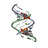
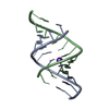
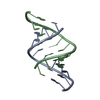
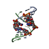
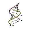
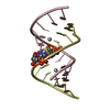
| Title | Crystal structure of an A-form RNA duplex containing three GU base pairs | ||||||
 Components Components |
| ||||||
 Keywords Keywords |  RNA / A-form RNA / ribose-zipper motif / GU base pair RNA / A-form RNA / ribose-zipper motif / GU base pair | ||||||
| Function / homology | RNA / RNA (> 10) Function and homology information Function and homology information | ||||||
| Method | X-RAY DIFFRACTION / SYNCHROTRON / MOLECULAR REPLACEMENT / molecular replacement / Resolution: 2.6 Å | ||||||
 Authors Authors | Kondo, J. / Dock-Bregeon, A.C. / Willkomm, D.K. / Hartmann, R.K. / Westhof, E. | ||||||
 Citation Citation | Journal: Acta Crystallogr.,Sect.F / Year: 2013 Title: Structure of an A-form RNA duplex obtained by degradation of 6S RNA in a crystallization droplet Authors: Kondo, J. / Dock-Bregeon, A.C. / Willkomm, D.K. / Hartmann, R.K. / Westhof, E. | ||||||
| History |
|
- Structure visualization
| Structure viewer | Molecule: MolmilJmol/JSmol |
|---|
- Downloads & links
-Download
| PDBx/mmCIF format | 4jrt.cif.gz | 23.2 KB | Display | PDBx/mmCIF format |
|---|---|---|---|---|
| PDB format | pdb4jrt.ent.gz | 14.7 KB | Display | PDB format |
| PDBx/mmJSON format | 4jrt.json.gz | Tree view | PDBx/mmJSON format | |
| Others |  Other downloads Other downloads |
-Validation report
| Arichive directory | https://data.pdbj.org/pub/pdb/validation_reports/jr/4jrtftp://data.pdbj.org/pub/pdb/validation_reports/jr/4jrt | HTTPS FTP |
|---|
-Related structure data
| Related structure data |  353dS S: Starting model for refinement |
|---|---|
| Similar structure data |

-Links
 PDBj
PDBj


- Assembly
| Deposited unit | 
| ||||||||
|---|---|---|---|---|---|---|---|---|---|
| 1 |
| ||||||||
| Unit cell |
|
-Components
| #1: RNA chain | Mass: 3979.408 Da / Num. of mol.: 1 / Source method: obtained synthetically / Details: in vitro transcription |
|---|---|
| #2: RNA chain | Mass: 3723.264 Da / Num. of mol.: 1 / Source method: obtained synthetically / Details: in vitro transcription |
| #3: Water | ChemComp-HOH / Water Mass: 18.015 Da / Num. of mol.: 23 / Source method: isolated from a natural source / Formula: H2O Mass: 18.015 Da / Num. of mol.: 23 / Source method: isolated from a natural source / Formula: H2O |
| Nonpolymer details | HOH (A 111) AND HOH (A 112) ARE CLOSED TO SYMMETRY-RELATED RNA MOLECULE. |
-Experimental details
-Experiment
| Experiment | Method: X-RAY DIFFRACTION / Number of used crystals: 1 |
|---|
- Sample preparation
| Crystal | Density Matthews: 2.51 Å3/Da / Density % sol: 50.92 % |
|---|---|
| Crystal grow | Temperature: 310 K / Method: vapor diffusion / pH: 7 Details: Sodium Cacodylate, Lithium acetate, PEG3350, pH 7.0, vapor diffusion, temperature 310K |
-Data collection
| Diffraction | Mean temperature: 100 K |
|---|---|
| Diffraction source | Source: SYNCHROTRON / Site: ESRF  / Beamline: ID23-1 / Wavelength: 1.0723 Å / Beamline: ID23-1 / Wavelength: 1.0723 Å |
| Detector | Detector: CCD / Date: Jul 4, 2009 |
| Radiation | Protocol: SINGLE WAVELENGTH / Monochromatic (M) / Laue (L): M / Scattering type: x-ray |
| Radiation wavelength | Wavelength: 1.0723 Å / Relative weight: 1 |
| Reflection | Resolution: 2.6→20.7 Å / Num. obs: 2330 |
-Phasing
| Phasing | Method: molecular replacement |
|---|
- Processing
| Software |
| |||||||||||||||||||||||||||||||||||||||||||||||||||||||||||||||||||||||||||||
|---|---|---|---|---|---|---|---|---|---|---|---|---|---|---|---|---|---|---|---|---|---|---|---|---|---|---|---|---|---|---|---|---|---|---|---|---|---|---|---|---|---|---|---|---|---|---|---|---|---|---|---|---|---|---|---|---|---|---|---|---|---|---|---|---|---|---|---|---|---|---|---|---|---|---|---|---|---|---|
| Refinement | Method to determine structure: MOLECULAR REPLACEMENT Starting model: PDB ENTRY 353D Resolution: 2.6→20.7 Å / Occupancy max: 1 / Occupancy min: 1 / σ(F): 0
| |||||||||||||||||||||||||||||||||||||||||||||||||||||||||||||||||||||||||||||
| Solvent computation | Bsol: 41.7378 Å2 | |||||||||||||||||||||||||||||||||||||||||||||||||||||||||||||||||||||||||||||
| Displacement parameters | Biso max: 113.1 Å2 / Biso mean: 76.1548 Å2 / Biso min: 41.68 Å2
| |||||||||||||||||||||||||||||||||||||||||||||||||||||||||||||||||||||||||||||
| Refinement step | Cycle: LAST / Resolution: 2.6→20.7 Å
| |||||||||||||||||||||||||||||||||||||||||||||||||||||||||||||||||||||||||||||
| Refine LS restraints |
| |||||||||||||||||||||||||||||||||||||||||||||||||||||||||||||||||||||||||||||
| LS refinement shell | Refine-ID: X-RAY DIFFRACTION / Total num. of bins used: 10
| |||||||||||||||||||||||||||||||||||||||||||||||||||||||||||||||||||||||||||||
| Xplor file |
|
