 Movie
Movie Controller

[English] 日本語
 Yorodumi
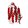
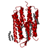
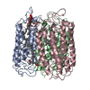
- PDB-4jr8: Crystal structure of cruxrhodopsin-3 from Haloarcula vallismortis... -
+ Open data

- Basic information
| Entry | Database: PDB / ID: 4jr8 | ||||||
|---|---|---|---|---|---|---|---|
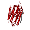
| Title | Crystal structure of cruxrhodopsin-3 from Haloarcula vallismortis at 2.3 angstrom resolution | ||||||
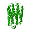
 Components Components | Cruxrhodopsin-3 | ||||||
 Keywords Keywords |  PROTON TRANSPORT / protein-bacteioruberin complex / seven transmembrane alpha helices / light-driven proton pump / Membrane PROTON TRANSPORT / protein-bacteioruberin complex / seven transmembrane alpha helices / light-driven proton pump / Membrane | ||||||
| Function / homology |  Function and homology informationphotoreceptor activity / phototransduction / proton transmembrane transport / monoatomic ion channel activity / plasma membrane Function and homology informationphotoreceptor activity / phototransduction / proton transmembrane transport / monoatomic ion channel activity / plasma membraneSimilarity search - Function | ||||||
| Biological species |  Haloarcula vallismortis (Halophile) Haloarcula vallismortis (Halophile) | ||||||
| Method | X-RAY DIFFRACTION / SYNCHROTRON / MOLECULAR REPLACEMENT / Resolution: 2.3 Å | ||||||
 Authors Authors | Kouyama, T. / Chan, S.K. | ||||||
 Citation Citation | Journal: Plos One / Year: 2014 Title: Crystal structure of Cruxrhodopsin-3 from Haloarcula vallismortis Authors: Chan, S.K. / Kitajima-Ihara, T. / Fujii, R. / Gotoh, T. / Murakami, M. / Ihara, K. / Kouyama, T. | ||||||
| History |
|
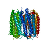
- Structure visualization
| Structure viewer | Molecule: MolmilJmol/JSmol |
|---|
- Downloads & links
-Download
| PDBx/mmCIF format | 4jr8.cif.gz | 60.1 KB | Display | PDBx/mmCIF format |
|---|---|---|---|---|
| PDB format | pdb4jr8.ent.gz | 42.6 KB | Display | PDB format |
| PDBx/mmJSON format | 4jr8.json.gz | Tree view | PDBx/mmJSON format | |
| Others |  Other downloads Other downloads |
-Validation report
| Arichive directory | https://data.pdbj.org/pub/pdb/validation_reports/jr/4jr8ftp://data.pdbj.org/pub/pdb/validation_reports/jr/4jr8 | HTTPS FTP |
|---|
-Related structure data
| Related structure data |  4l35C 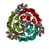 1iw6S S: Starting model for refinement C: citing same article ( |
|---|---|
| Similar structure data |
-Links
 PDBj
PDBj


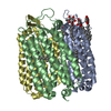
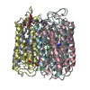
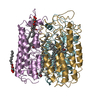
- Assembly
| Deposited unit | 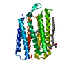
| ||||||||
|---|---|---|---|---|---|---|---|---|---|
| 1 | 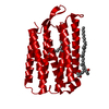
| ||||||||
| Unit cell |
|
-Components
| #1: Protein | Mass: 26867.229 Da / Num. of mol.: 1 Source method: isolated from a genetically manipulated source Source: (gene. exp.) Haloarcula vallismortis (Halophile) / Gene: cop3 / Production host: Halobacterium salinarum (Halophile) / References: UniProt: P94854 |
|---|---|
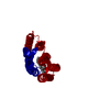
| #2: Chemical | ChemComp-RET / Retinal  Mass: 284.436 Da / Num. of mol.: 1 / Source method: obtained synthetically / Formula: C20H28O Mass: 284.436 Da / Num. of mol.: 1 / Source method: obtained synthetically / Formula: C20H28O |
| #3: Chemical | ChemComp-22B / Halobacterium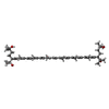  Mass: 741.136 Da / Num. of mol.: 1 / Source method: obtained synthetically / Formula: C50H76O4 Mass: 741.136 Da / Num. of mol.: 1 / Source method: obtained synthetically / Formula: C50H76O4 |
| #4: Water | ChemComp-HOH / Water Mass: 18.015 Da / Num. of mol.: 14 / Source method: isolated from a natural source / Formula: H2O Mass: 18.015 Da / Num. of mol.: 14 / Source method: isolated from a natural source / Formula: H2O |
-Experimental details
-Experiment
| Experiment | Method: X-RAY DIFFRACTION / Number of used crystals: 1 |
|---|
- Sample preparation
| Crystal | Density Matthews: 3.65 Å3/Da / Density % sol: 66.29 % |
|---|---|
| Crystal grow | Temperature: 288 K / Method: vapor diffusion, sitting drop / pH: 4 Details: 2.1M ammonium sulfate, 0.1M sodium citrate, pH 4.0, VAPOR DIFFUSION, SITTING DROP, temperature 288K |
-Data collection
| Diffraction | Mean temperature: 100 K |
|---|---|
| Diffraction source | Source: SYNCHROTRON / Site: SPring-8  / Beamline: BL38B1 / Wavelength: 1 Å / Beamline: BL38B1 / Wavelength: 1 Å |
| Detector | Type: ADSC QUANTUM 315 / Detector: CCD / Date: Nov 7, 2012 |
| Radiation | Monochromator: Si (111) double crystal monochromator / Protocol: SINGLE WAVELENGTH / Monochromatic (M) / Laue (L): M / Scattering type: x-ray |
| Radiation wavelength | Wavelength: 1 Å / Relative weight: 1 |
| Reflection | Resolution: 2.3→45.9 Å / Num. all: 17545 / Num. obs: 15610 / % possible obs: 90 % / Observed criterion σ(F): 2.3 / Observed criterion σ(I): 2.3 / Redundancy: 5.9 % / Biso Wilson estimate: 26 Å2 / Rmerge(I) obs: 0.074 / Rsym value: 0.074 / Net I/σ(I): 21.2 |
| Reflection shell | Resolution: 2.3→2.42 Å / Redundancy: 5.9 % / Rmerge(I) obs: 0.448 / Mean I/σ(I) obs: 4.4 / Num. unique all: 2299 / Rsym value: 0.448 / % possible all: 92.4 |
- Processing
| Software |
| ||||||||||||||||||||
|---|---|---|---|---|---|---|---|---|---|---|---|---|---|---|---|---|---|---|---|---|---|
| Refinement | Method to determine structure: MOLECULAR REPLACEMENT Starting model: PDB ENTRY 1iw6 Resolution: 2.3→15 Å / Isotropic thermal model: Isotropic / σ(F): 0 / σ(I): 0 / Stereochemistry target values: Engh & Huber
| ||||||||||||||||||||
| Displacement parameters | Biso mean: 33.2 Å2
| ||||||||||||||||||||
| Refine analyze |
| ||||||||||||||||||||
| Refinement step | Cycle: LAST / Resolution: 2.3→15 Å
| ||||||||||||||||||||
| Refine LS restraints |
|
