 Movie
Movie Controller


+ Open data

- Basic information
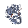
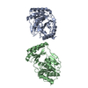
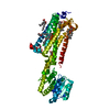
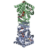
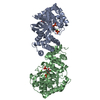
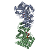
| Entry | Database: PDB / ID: 4jhj | ||||||
|---|---|---|---|---|---|---|---|
| Title | Crystal structure of Danio rerio Slip1 in complex with Dbp5 | ||||||
 Components Components |
| ||||||
 Keywords Keywords |  TRANSLATION / DEAD-BOX HELICASE / MRNA EXPORT / RECA-LIKE / RNA-DEPENDENT ATPASE / DDX19 / DEAD-BOX PROTEIN 19B / MRNA TRANSPORT / ATP BINDING / HELICASE / HYDROLASE / NUCLEOTIDE BINDING / RNA BINDING / TRANSPORT HISTONE MRNA PROCESSING / 3'-UTR / 3'-PROCESSING / NUP214 TRANSLATION / DEAD-BOX HELICASE / MRNA EXPORT / RECA-LIKE / RNA-DEPENDENT ATPASE / DDX19 / DEAD-BOX PROTEIN 19B / MRNA TRANSPORT / ATP BINDING / HELICASE / HYDROLASE / NUCLEOTIDE BINDING / RNA BINDING / TRANSPORT HISTONE MRNA PROCESSING / 3'-UTR / 3'-PROCESSING / NUP214 | ||||||
| Function / homology |  Function and homology information Function and homology informationhistone mRNA stem-loop binding complex / cap-dependent translational initiation / translation activator activity / regulation of translational initiation / poly(A)+ mRNA export from nucleus / cytoplasmic stress granule / RNA helicase activity / RNA helicase / RNA binding / ATP binding ...histone mRNA stem-loop binding complex / cap-dependent translational initiation / translation activator activity / regulation of translational initiation / poly(A)+ mRNA export from nucleus / cytoplasmic stress granule / RNA helicase activity / RNA helicase / RNA binding / ATP binding / identical protein binding / nucleus / cytosol / cytoplasmSimilarity search - Function | ||||||
| Biological species |  Danio rerio (zebrafish) Danio rerio (zebrafish) | ||||||
| Method | X-RAY DIFFRACTION / SYNCHROTRON / MOLECULAR REPLACEMENT / Resolution: 3.25 Å | ||||||
 Authors Authors | Von Moeller, H. / Conti, E. | ||||||
 Citation Citation | Journal: Nucleic Acids Res. / Year: 2013 Title: Structural and biochemical studies of SLIP1-SLBP identify DBP5 and eIF3g as SLIP1-binding proteins. Authors: von Moeller, H. / Lerner, R. / Ricciardi, A. / Basquin, C. / Marzluff, W.F. / Conti, E. | ||||||
| History |
|
- Structure visualization
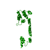
| Structure viewer | Molecule: MolmilJmol/JSmol |
|---|
- Downloads & links
-Download
| PDBx/mmCIF format | 4jhj.cif.gz | 102 KB | Display | PDBx/mmCIF format |
|---|---|---|---|---|
| PDB format | pdb4jhj.ent.gz | 79.2 KB | Display | PDB format |
| PDBx/mmJSON format | 4jhj.json.gz | Tree view | PDBx/mmJSON format | |
| Others |  Other downloads Other downloads |
-Validation report
| Arichive directory | https://data.pdbj.org/pub/pdb/validation_reports/jh/4jhjftp://data.pdbj.org/pub/pdb/validation_reports/jh/4jhj | HTTPS FTP |
|---|
-Related structure data
| Related structure data | 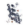 4jhkC 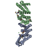 2i2oS S: Starting model for refinement C: citing same article ( |
|---|---|
| Similar structure data |


-Links
 PDBj
PDBj

- Assembly
| Deposited unit | 
| ||||||||||||||||||||||||||||||||||||||||||||||||||||||||||||||||||||
|---|---|---|---|---|---|---|---|---|---|---|---|---|---|---|---|---|---|---|---|---|---|---|---|---|---|---|---|---|---|---|---|---|---|---|---|---|---|---|---|---|---|---|---|---|---|---|---|---|---|---|---|---|---|---|---|---|---|---|---|---|---|---|---|---|---|---|---|---|---|
| 1 | 
| ||||||||||||||||||||||||||||||||||||||||||||||||||||||||||||||||||||
| 2 |
| ||||||||||||||||||||||||||||||||||||||||||||||||||||||||||||||||||||
| Unit cell |
| ||||||||||||||||||||||||||||||||||||||||||||||||||||||||||||||||||||
| Noncrystallographic symmetry (NCS) | NCS domain:
NCS domain segments: Component-ID: 1 / Refine code: 6
NCS ensembles :
|
-Components
| #1: Protein | Mass: 25794.500 Da / Num. of mol.: 2 / Fragment: UNP RESIDUES 1-222 Source method: isolated from a genetically manipulated source Source: (gene. exp.) Danio rerio (zebrafish) / Gene: mif4gdb, zgc:110826 / Production host:  ESCHERICHIA COLI (E. coli) / Strain (production host): BL21 / References: UniProt: Q5EAQ1 ESCHERICHIA COLI (E. coli) / Strain (production host): BL21 / References: UniProt: Q5EAQ1#2: Protein/peptide | Mass: 3495.730 Da / Num. of mol.: 2 / Fragment: UNP RESIDUES 1-32 Source method: isolated from a genetically manipulated source Source: (gene. exp.) Danio rerio (zebrafish) / Gene: ddx19 / Production host: ESCHERICHIA COLI (E. coli) / Strain (production host): BL21References: UniProt: Q7ZU28, Hydrolases; Acting on acid anhydrides; In phosphorus-containing anhydrides#3: Water | ChemComp-HOH / | Water Mass: 18.015 Da / Num. of mol.: 11 / Source method: isolated from a natural source / Formula: H2O Mass: 18.015 Da / Num. of mol.: 11 / Source method: isolated from a natural source / Formula: H2O |
|---|
-Experimental details
-Experiment
| Experiment | Method: X-RAY DIFFRACTION / Number of used crystals: 1 |
|---|
- Sample preparation
| Crystal | Density Matthews: 4.73 Å3/Da / Density % sol: 73.98 % |
|---|---|
| Crystal grow | Temperature: 293 K / Method: vapor diffusion, sitting drop / pH: 6 Details: 0.05 M MES pH 6.0, 18% (w/v) PEG 8000, 0.2 M Ca-Acetate, VAPOR DIFFUSION, SITTING DROP, temperature 293K |
-Data collection
| Diffraction | Mean temperature: 100 K |
|---|---|
| Diffraction source | Source: SYNCHROTRON / Site: SLS  / Beamline: X06DA / Beamline: X06DA |
| Detector | Type: MARMOSAIC 225 mm CCD / Detector: CCD / Date: Mar 22, 2010 |
| Radiation | Protocol: SINGLE WAVELENGTH / Monochromatic (M) / Laue (L): M / Scattering type: x-ray |
| Radiation wavelength | Relative weight: 1 |
| Reflection | Resolution: 3.25→86.13 Å / Num. obs: 16724 / % possible obs: 99.8 % / Observed criterion σ(I): 2 / Redundancy: 4.5 % / Rsym value: 0.083 / Net I/σ(I): 16.34 |
| Reflection shell | Resolution: 3.25→3.45 Å / Redundancy: 4.6 % / Mean I/σ(I) obs: 2.3 / Rsym value: 0.68 / % possible all: 99.9 |
- Processing
| Software |
| ||||||||||||||||||||||||||||||||||||||||||||||||||||||||||||||||||||||||||||||||||||||||||||||||||||||||||||||||||||||||||||||||||||||||||||||||||||||||||||||||||||||||||
|---|---|---|---|---|---|---|---|---|---|---|---|---|---|---|---|---|---|---|---|---|---|---|---|---|---|---|---|---|---|---|---|---|---|---|---|---|---|---|---|---|---|---|---|---|---|---|---|---|---|---|---|---|---|---|---|---|---|---|---|---|---|---|---|---|---|---|---|---|---|---|---|---|---|---|---|---|---|---|---|---|---|---|---|---|---|---|---|---|---|---|---|---|---|---|---|---|---|---|---|---|---|---|---|---|---|---|---|---|---|---|---|---|---|---|---|---|---|---|---|---|---|---|---|---|---|---|---|---|---|---|---|---|---|---|---|---|---|---|---|---|---|---|---|---|---|---|---|---|---|---|---|---|---|---|---|---|---|---|---|---|---|---|---|---|---|---|---|---|---|---|---|
| Refinement | Method to determine structure: MOLECULAR REPLACEMENT Starting model: 2I2O Resolution: 3.25→73.77 Å / Cor.coef. Fo:Fc: 0.931 / Cor.coef. Fo:Fc free: 0.912 / SU B: 25.36 / SU ML: 0.403 / Cross valid method: THROUGHOUT / ESU R Free: 0.446 / Stereochemistry target values: MAXIMUM LIKELIHOOD / Details: HYDROGENS HAVE BEEN ADDED IN THE RIDING POSITIONS
| ||||||||||||||||||||||||||||||||||||||||||||||||||||||||||||||||||||||||||||||||||||||||||||||||||||||||||||||||||||||||||||||||||||||||||||||||||||||||||||||||||||||||||
| Solvent computation | Ion probe radii: 0.8 Å / Shrinkage radii: 0.8 Å / VDW probe radii: 1.4 Å / Solvent model: MASK | ||||||||||||||||||||||||||||||||||||||||||||||||||||||||||||||||||||||||||||||||||||||||||||||||||||||||||||||||||||||||||||||||||||||||||||||||||||||||||||||||||||||||||
| Displacement parameters | Biso mean: 102.3 Å2
| ||||||||||||||||||||||||||||||||||||||||||||||||||||||||||||||||||||||||||||||||||||||||||||||||||||||||||||||||||||||||||||||||||||||||||||||||||||||||||||||||||||||||||
| Refinement step | Cycle: LAST / Resolution: 3.25→73.77 Å
| ||||||||||||||||||||||||||||||||||||||||||||||||||||||||||||||||||||||||||||||||||||||||||||||||||||||||||||||||||||||||||||||||||||||||||||||||||||||||||||||||||||||||||
| Refine LS restraints |
| ||||||||||||||||||||||||||||||||||||||||||||||||||||||||||||||||||||||||||||||||||||||||||||||||||||||||||||||||||||||||||||||||||||||||||||||||||||||||||||||||||||||||||
| Refine LS restraints NCS | Dom-ID: 1 / Refine-ID: X-RAY DIFFRACTION
| ||||||||||||||||||||||||||||||||||||||||||||||||||||||||||||||||||||||||||||||||||||||||||||||||||||||||||||||||||||||||||||||||||||||||||||||||||||||||||||||||||||||||||
| LS refinement shell | Resolution: 3.25→3.33 Å / Total num. of bins used: 20
|
