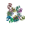
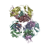
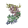
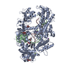
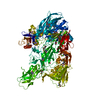
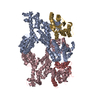
- PDB-4jcv: Crystal structure of the RecOR complex in an open conformation -
+
Open data
ID or keywords:
Loading...
-
Basic information
Entry
Database: PDB / ID: 4jcv
Title
Crystal structure of the RecOR complex in an open conformation
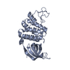
Components
DNA repair protein RecO
Recombination protein RecRGenetic recombination
Keywords
RECOMBINATION / HOMOLOGOUS RECOMBINATION / RECFOR PATHWAY / DNA BINDING / ZINC-FINGER / DNA DAMAGE / DNA REPAIR / RECR / RECO / RECOR
Function / homology
Function and homology information
bacterial nucleoid / double-strand break repair / DNA recombination / DNA binding / metal ion binding Similarity search - Function
Recombination protein O, zinc-binding domain / Recombination protein O, C-terminal domain / Erythroid Transcription Factor GATA-1; Chain A / Recombination protein O, C-terminal / Recombination protein O, RecO / DNA replication/recombination mediator RecO, N-terminal / Recombination protein O C terminal / Recombination protein O N terminal / RecR, C-terminal / RecR, helix-hairpin-helix ...Recombination protein O, zinc-binding domain / Recombination protein O, C-terminal domain / Erythroid Transcription Factor GATA-1; Chain A / Recombination protein O, C-terminal / Recombination protein O, RecO / DNA replication/recombination mediator RecO, N-terminal / Recombination protein O C terminal / Recombination protein O N terminal / RecR, C-terminal / RecR, helix-hairpin-helix / DNA recombination protein RecR / Recombination protein RecR, conserved site / Recombination protein RecR / RecR, TOPRIM domain / RecR, Cys4-zinc finger motif / RecR protein signature. / Toprim domain / Dna Topoisomerase Vi A Subunit; Chain: A, domain 2 / Dna Topoisomerase Vi A Subunit; Chain: A, domain 2 - #10 / ARFGAP/RecO-like zinc finger / de novo design (two linked rop proteins) / TOPRIM / Toprim domain / Other non-globular / Toprim domain profile. / TOPRIM domain / Helix-hairpin-helix DNA-binding motif, class 1 / Helix-hairpin-helix DNA-binding motif class 1 / Nucleic acid-binding proteins / Special / OB fold (Dihydrolipoamide Acetyltransferase, E2P) / Nucleic acid-binding, OB-fold / Up-down Bundle / Beta Barrel / 3-Layer(aba) Sandwich / Mainly Beta / Mainly Alpha / Alpha Beta Similarity search - Domain/homology
A: Recombination protein RecR B: Recombination protein RecR C: Recombination protein RecR D: Recombination protein RecR E: DNA repair protein RecO F: DNA repair protein RecO hetero molecules
Resolution: 3.34→83.07 Å / Cor.coef. Fo:Fc: 0.917 / Cor.coef. Fo:Fc free: 0.903 / SU B: 73.357 / SU ML: 0.552 / Cross valid method: THROUGHOUT / σ(I): 2 / ESU R Free: 0.611 / Stereochemistry target values: MAXIMUM LIKELIHOOD / Details: HYDROGENS HAVE BEEN USED IF PRESENT IN THE INPUT
Rfactor
Num. reflection
% reflection
Selection details
Rfree
0.2781
1212
5 %
RANDOM
Rwork
0.24016
-
-
-
obs
0.24203
22815
94.21 %
-
all
-
24029
-
-
Solvent computation
Ion probe radii: 0.8 Å / Shrinkage radii: 0.8 Å / VDW probe radii: 1.2 Å / Solvent model: MASK
Displacement parameters
Biso mean: 113.5 Å2
Baniso -1
Baniso -2
Baniso -3
1-
-1.53 Å2
2.68 Å2
3.19 Å2
2-
-
3.58 Å2
4.7 Å2
3-
-
-
3.89 Å2
Refinement step
Cycle: LAST / Resolution: 3.34→83.07 Å
Protein
Nucleic acid
Ligand
Solvent
Total
Num. atoms
9156
0
6
0
9162
Refine LS restraints
Refine-ID
Type
Dev ideal
Dev ideal target
Number
X-RAY DIFFRACTION
r_bond_refined_d
0.01
0.019
9343
X-RAY DIFFRACTION
r_bond_other_d
X-RAY DIFFRACTION
r_angle_refined_deg
1.329
1.992
12713
X-RAY DIFFRACTION
r_angle_other_deg
X-RAY DIFFRACTION
r_dihedral_angle_1_deg
7.16
5
1196
X-RAY DIFFRACTION
r_dihedral_angle_2_deg
35.038
22.872
383
X-RAY DIFFRACTION
r_dihedral_angle_3_deg
16.707
15
1513
X-RAY DIFFRACTION
r_dihedral_angle_4_deg
16.314
15
87
X-RAY DIFFRACTION
r_chiral_restr
0.084
0.2
1473
X-RAY DIFFRACTION
r_gen_planes_refined
0.01
0.022
7080
X-RAY DIFFRACTION
r_gen_planes_other
X-RAY DIFFRACTION
r_nbd_refined
X-RAY DIFFRACTION
r_nbd_other
X-RAY DIFFRACTION
r_nbtor_refined
X-RAY DIFFRACTION
r_nbtor_other
X-RAY DIFFRACTION
r_xyhbond_nbd_refined
X-RAY DIFFRACTION
r_xyhbond_nbd_other
X-RAY DIFFRACTION
r_metal_ion_refined
X-RAY DIFFRACTION
r_metal_ion_other
X-RAY DIFFRACTION
r_symmetry_vdw_refined
X-RAY DIFFRACTION
r_symmetry_vdw_other
X-RAY DIFFRACTION
r_symmetry_hbond_refined
X-RAY DIFFRACTION
r_symmetry_hbond_other
X-RAY DIFFRACTION
r_symmetry_metal_ion_refined
X-RAY DIFFRACTION
r_symmetry_metal_ion_other
X-RAY DIFFRACTION
r_mcbond_it
X-RAY DIFFRACTION
r_mcbond_other
X-RAY DIFFRACTION
r_mcangle_it
X-RAY DIFFRACTION
r_scbond_it
X-RAY DIFFRACTION
r_scangle_it
X-RAY DIFFRACTION
r_rigid_bond_restr
X-RAY DIFFRACTION
r_sphericity_free
X-RAY DIFFRACTION
r_sphericity_bonded
LS refinement shell
Resolution: 3.34→3.427 Å / Total num. of bins used: 20
Rfactor
Num. reflection
% reflection
Rfree
0.356
92
-
Rwork
0.316
1709
-
obs
-
-
96.26 %
Refinement TLS params.
Method: refined / Refine-ID: X-RAY DIFFRACTION
ID
L11 (°2)
L12 (°2)
L13 (°2)
L22 (°2)
L23 (°2)
L33 (°2)
S11 (Å °)
S12 (Å °)
S13 (Å °)
S21 (Å °)
S22 (Å °)
S23 (Å °)
S31 (Å °)
S32 (Å °)
S33 (Å °)
T11 (Å2)
T12 (Å2)
T13 (Å2)
T22 (Å2)
T23 (Å2)
T33 (Å2)
Origin x (Å)
Origin y (Å)
Origin z (Å)
1
5.161
-2.463
0.4207
3.7345
-0.2687
1.2457
-0.0004
-0.5687
-0.1837
-0.3116
0.2154
0.1988
0.1335
-0.0495
-0.215
0.2024
-0.1044
0.0184
0.1972
0.0594
0.173
-46.3221
33.3972
22.1079
2
4.4038
-0.8359
1.6536
1.419
-0.8307
2.308
-0.0888
0.0565
0.5255
-0.2218
-0.0498
-0.2186
0.0568
0.1398
0.1386
0.2397
-0.0706
0.1052
0.1096
0.0465
0.2232
-77.026
53.7121
10.2891
3
3.7197
-2.0551
-0.1959
5.6246
0.7426
2.0324
0.5583
0.8261
-0.6427
-0.8935
-0.282
0.1236
0.8144
-0.3202
-0.2763
0.8307
0.0354
-0.0375
0.7857
0.1649
0.4636
-77.1639
36.6008
-21.352
4
3.2706
0.4537
2.0514
1.2512
0.2977
5.0018
0.2315
0.7422
-0.5213
-0.7969
0.0611
0.0059
0.2372
0.0625
-0.2925
0.8998
-0.0132
-0.1199
0.2826
-0.0366
0.9598
-51.4404
9.0352
-4.2263
5
4.1952
0.9018
-0.1408
3.532
-1.8479
6.549
-0.0585
-0.675
0.0928
0.7705
0.098
-0.3537
-0.2228
0.6746
-0.0395
0.3771
0.1242
0.0491
0.8828
0.0387
0.3042
-54.5144
58.1699
-24.7346
6
4.0467
0.7133
-0.6258
9.6283
-1.0297
2.171
-0.3372
-0.6211
1.0697
0.5684
0.1221
0.5391
-0.7047
-0.5589
0.2152
0.4551
0.125
-0.1855
0.4906
-0.2819
0.9437
-71.7872
-0.9854
17.4729
Refinement TLS group
ID
Refine-ID
Refine TLS-ID
Auth asym-ID
Auth seq-ID
1
X-RAY DIFFRACTION
1
A
2 - 197
2
X-RAY DIFFRACTION
2
B
2 - 197
3
X-RAY DIFFRACTION
3
C
9 - 199
4
X-RAY DIFFRACTION
4
D
4 - 199
5
X-RAY DIFFRACTION
5
E
3 - 226
6
X-RAY DIFFRACTION
6
F
5 - 227
+
About Yorodumi
-
News
-
Feb 9, 2022. New format data for meta-information of EMDB entries
New format data for meta-information of EMDB entries
Version 3 of the EMDB header file is now the official format.
The previous official version 1.9 will be removed from the archive.
In the structure databanks used in Yorodumi, some data are registered as the other names, "COVID-19 virus" and "2019-nCoV". Here are the details of the virus and the list of structure data.
Jan 31, 2019. EMDB accession codes are about to change! (news from PDBe EMDB page)
EMDB accession codes are about to change! (news from PDBe EMDB page)
The allocation of 4 digits for EMDB accession codes will soon come to an end. Whilst these codes will remain in use, new EMDB accession codes will include an additional digit and will expand incrementally as the available range of codes is exhausted. The current 4-digit format prefixed with “EMD-” (i.e. EMD-XXXX) will advance to a 5-digit format (i.e. EMD-XXXXX), and so on. It is currently estimated that the 4-digit codes will be depleted around Spring 2019, at which point the 5-digit format will come into force.
The EM Navigator/Yorodumi systems omit the EMD- prefix.
Related info.:Q: What is EMD? / ID/Accession-code notation in Yorodumi/EM Navigator
Yorodumi is a browser for structure data from EMDB, PDB, SASBDB, etc.
This page is also the successor to EM Navigator detail page, and also detail information page/front-end page for Omokage search.
The word "yorodu" (or yorozu) is an old Japanese word meaning "ten thousand". "mi" (miru) is to see.
Related info.:EMDB / PDB / SASBDB / Comparison of 3 databanks / Yorodumi Search / Aug 31, 2016. New EM Navigator & Yorodumi / Yorodumi Papers / Jmol/JSmol / Function and homology information / Changes in new EM Navigator and Yorodumi
 Movie
Movie Controller


 Open data
 Basic information
 Components
 Keywords
 RECOMBINATION /
  Function and homology information
 Authors
 Citation
 Structure visualization
 Downloads & links
 Other downloads
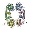

 PDBj
PDBj


 Assembly

 Mass: 65.409 Da / Num. of mol.: 6 / Source method: obtained synthetically / Formula: Zn
 Sample preparation
 / Beamline: ID14-4 / Wavelength: 0.9793 Å
 Processing