 Movie
Movie Controller

[English] 日本語
 Yorodumi
- PDB-4jcu: Crystal structure of a 5-carboxymethyl-2-hydroxymuconate isomeras... -
+ Open data

- Basic information
| Entry | Database: PDB / ID: 4jcu | ||||||
|---|---|---|---|---|---|---|---|
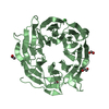
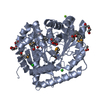
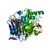
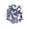
| Title | Crystal structure of a 5-carboxymethyl-2-hydroxymuconate isomerase from Deinococcus radiodurans R1 | ||||||
 Components Components | 5-carboxymethyl-2-hydroxymuconate isomerase | ||||||
 Keywords Keywords |  ISOMERASE / Structural Genomics / PSI-Biology / New York Structural Genomics Research Consortium / NYSGRC / alpha/beta / Triangular trimer assembly / PROTEIN STRUCTURE INITIATIVE / 5-carboxymethyl-2-hydroxymuconate ISOMERASE / Structural Genomics / PSI-Biology / New York Structural Genomics Research Consortium / NYSGRC / alpha/beta / Triangular trimer assembly / PROTEIN STRUCTURE INITIATIVE / 5-carboxymethyl-2-hydroxymuconate | ||||||
| Function / homology |  Function and homology information Function and homology information | ||||||
| Biological species |  Deinococcus radiodurans (radioresistant) Deinococcus radiodurans (radioresistant) | ||||||
| Method | X-RAY DIFFRACTION / SYNCHROTRON / SAD / Resolution: 2.4 Å | ||||||
 Authors Authors | Kumaran, D. / Chamala, S. / Evans, B. / Foti, R. / Gizzi, A. / Hillerich, B. / Kar, A. / Lafleur, J. / Seidel, R. / Villigas, G. ...Kumaran, D. / Chamala, S. / Evans, B. / Foti, R. / Gizzi, A. / Hillerich, B. / Kar, A. / Lafleur, J. / Seidel, R. / Villigas, G. / Zencheck, W. / Al Obaidi, N. / Almo, S.C. / Swaminathan, S. / New York Structural Genomics Research Consortium (NYSGRC) | ||||||
 Citation Citation | Journal: To be Published Title: Crystal structure of a 5-carboxymethyl-2-hydroxymuconate isomerase from Deinococcus radiodurans R1 Authors: Kumaran, D. / Almo, S.C. / Swaminathan, S. | ||||||
| History |
|
- Structure visualization
| Structure viewer | Molecule: MolmilJmol/JSmol |
|---|
- Downloads & links
-Download
| PDBx/mmCIF format | 4jcu.cif.gz | 86.7 KB | Display | PDBx/mmCIF format |
|---|---|---|---|---|
| PDB format | pdb4jcu.ent.gz | 71.5 KB | Display | PDB format |
| PDBx/mmJSON format | 4jcu.json.gz | Tree view | PDBx/mmJSON format | |
| Others |  Other downloads Other downloads |
-Validation report
| Arichive directory | https://data.pdbj.org/pub/pdb/validation_reports/jc/4jcuftp://data.pdbj.org/pub/pdb/validation_reports/jc/4jcu | HTTPS FTP |
|---|
-Related structure data
| Similar structure data | |
|---|---|
| Other databases |


-Links
 PDBj
PDBj- Assembly
| Deposited unit | 
| ||||||||
|---|---|---|---|---|---|---|---|---|---|
| 1 |
| ||||||||
| Unit cell |
|
-Components
| #1: Protein | Mass: 17071.732 Da / Num. of mol.: 3 Source method: isolated from a genetically manipulated source Source: (gene. exp.) Deinococcus radiodurans (radioresistant)Strain: R1 / Gene: DR_A0227 / Plasmid: PET3A / Production host: Escherichia coli (E. coli) / Strain (production host): BL21(DE3) / References: UniProt: Q9RYT1#2: Water | ChemComp-HOH / | Water Mass: 18.015 Da / Num. of mol.: 39 / Source method: isolated from a natural source / Formula: H2O Mass: 18.015 Da / Num. of mol.: 39 / Source method: isolated from a natural source / Formula: H2O |
|---|
-Experimental details
-Experiment
| Experiment | Method: X-RAY DIFFRACTION / Number of used crystals: 1 |
|---|
- Sample preparation
| Crystal | Density Matthews: 2.66 Å3/Da / Density % sol: 53.85 % |
|---|---|
| Crystal grow | Temperature: 298 K / Method: vapor diffusion, sitting drop / pH: 4.5 Details: 0.2M Ammonium acetate, 0.1M Sodium acetate, 25% PEG 4000, 5% Trimethylamine N-oxide dihydrate, pH 4.5, VAPOR DIFFUSION, SITTING DROP, temperature 298K |
-Data collection
| Diffraction | Mean temperature: 100 K |
|---|---|
| Diffraction source | Source: SYNCHROTRON / Site: NSLS  / Beamline: X29A / Wavelength: 0.979 Å / Beamline: X29A / Wavelength: 0.979 Å |
| Detector | Type: ADSC QUANTUM 315 / Detector: CCD / Date: Feb 21, 2013 / Details: Mirrors |
| Radiation | Monochromator: Si III / Protocol: SINGLE WAVELENGTH / Monochromatic (M) / Laue (L): M / Scattering type: x-ray |
| Radiation wavelength | Wavelength: 0.979 Å / Relative weight: 1 |
| Reflection | Resolution: 2.4→50 Å / Num. all: 21677 / Num. obs: 21677 / % possible obs: 99.8 % / Observed criterion σ(F): 0 / Observed criterion σ(I): 0 / Redundancy: 14.6 % / Rmerge(I) obs: 0.083 / Net I/σ(I): 14.8 |
| Reflection shell | Resolution: 2.4→2.47 Å / Redundancy: 12.2 % / Rmerge(I) obs: 0.448 / Mean I/σ(I) obs: 2 / Num. unique all: 1749 / % possible all: 99 |
- Processing
| Software |
| ||||||||||||||||||||||||||||||||||||||||||||||||||||||||||||||||||||||||||||||||||||||||||||||||||||||||||||||||||||||||||||||||||||||||||||||||||||||||||||||||||||||||||
|---|---|---|---|---|---|---|---|---|---|---|---|---|---|---|---|---|---|---|---|---|---|---|---|---|---|---|---|---|---|---|---|---|---|---|---|---|---|---|---|---|---|---|---|---|---|---|---|---|---|---|---|---|---|---|---|---|---|---|---|---|---|---|---|---|---|---|---|---|---|---|---|---|---|---|---|---|---|---|---|---|---|---|---|---|---|---|---|---|---|---|---|---|---|---|---|---|---|---|---|---|---|---|---|---|---|---|---|---|---|---|---|---|---|---|---|---|---|---|---|---|---|---|---|---|---|---|---|---|---|---|---|---|---|---|---|---|---|---|---|---|---|---|---|---|---|---|---|---|---|---|---|---|---|---|---|---|---|---|---|---|---|---|---|---|---|---|---|---|---|---|---|
| Refinement | Method to determine structure: SAD / Resolution: 2.4→45.46 Å / Cor.coef. Fo:Fc: 0.95 / Cor.coef. Fo:Fc free: 0.916 / SU B: 8.082 / SU ML: 0.184 / Cross valid method: THROUGHOUT / σ(F): 0 / σ(I): 0 / ESU R: 0.319 / ESU R Free: 0.252 / Stereochemistry target values: MAXIMUM LIKELIHOOD / Details: HYDROGENS HAVE BEEN ADDED IN THE RIDING POSITIONS
| ||||||||||||||||||||||||||||||||||||||||||||||||||||||||||||||||||||||||||||||||||||||||||||||||||||||||||||||||||||||||||||||||||||||||||||||||||||||||||||||||||||||||||
| Solvent computation | Ion probe radii: 0.8 Å / Shrinkage radii: 0.8 Å / VDW probe radii: 1.2 Å / Solvent model: MASK | ||||||||||||||||||||||||||||||||||||||||||||||||||||||||||||||||||||||||||||||||||||||||||||||||||||||||||||||||||||||||||||||||||||||||||||||||||||||||||||||||||||||||||
| Displacement parameters | Biso mean: 44.276 Å2
| ||||||||||||||||||||||||||||||||||||||||||||||||||||||||||||||||||||||||||||||||||||||||||||||||||||||||||||||||||||||||||||||||||||||||||||||||||||||||||||||||||||||||||
| Refinement step | Cycle: LAST / Resolution: 2.4→45.46 Å
| ||||||||||||||||||||||||||||||||||||||||||||||||||||||||||||||||||||||||||||||||||||||||||||||||||||||||||||||||||||||||||||||||||||||||||||||||||||||||||||||||||||||||||
| Refine LS restraints |
| ||||||||||||||||||||||||||||||||||||||||||||||||||||||||||||||||||||||||||||||||||||||||||||||||||||||||||||||||||||||||||||||||||||||||||||||||||||||||||||||||||||||||||
| LS refinement shell | Resolution: 2.398→2.46 Å / Total num. of bins used: 20
|
