 Movie
Movie Controller

[English] 日本語
 Yorodumi
- PDB-4j3l: Tankyrase 2 in complex with 3-chloro-N-(2-methoxyethyl)-4-(4-meth... -
+ Open data

- Basic information
| Entry | Database: PDB / ID: 4j3l | ||||||
|---|---|---|---|---|---|---|---|
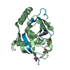
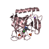
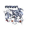
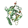
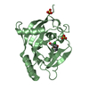
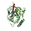
| Title | Tankyrase 2 in complex with 3-chloro-N-(2-methoxyethyl)-4-(4-methyl-2-oxo-1,2-dihydroquinolin-7-yl)benzamide | ||||||
 Components Components | Tankyrase-2 | ||||||
 Keywords Keywords | TRANSFERASE / ribosylation | ||||||
| Function / homology |  Function and homology information Function and homology informationXAV939 stabilizes AXIN / NAD+ ADP-ribosyltransferase / negative regulation of telomere maintenance via telomere lengthening / protein auto-ADP-ribosylation / protein localization to chromosome, telomeric region / protein poly-ADP-ribosylation / pericentriolar material / NAD+-protein ADP-ribosyltransferase activity / positive regulation of telomere capping / Transferases; Glycosyltransferases; Pentosyltransferases ...XAV939 stabilizes AXIN / NAD+ ADP-ribosyltransferase / negative regulation of telomere maintenance via telomere lengthening / protein auto-ADP-ribosylation / protein localization to chromosome, telomeric region / protein poly-ADP-ribosylation / pericentriolar material / NAD+-protein ADP-ribosyltransferase activity / positive regulation of telomere capping / Transferases; Glycosyltransferases; Pentosyltransferases / NAD+ ADP-ribosyltransferase activity / positive regulation of telomere maintenance via telomerase / nucleotidyltransferase activity / TCF dependent signaling in response to WNT / Degradation of AXIN / Wnt signaling pathway / Regulation of PTEN stability and activity / protein polyubiquitination / positive regulation of canonical Wnt signaling pathway / nuclear envelope / chromosome, telomeric region / Ub-specific processing proteases / Golgi membrane / perinuclear region of cytoplasm / enzyme binding / metal ion binding / nucleus / cytosol / cytoplasmSimilarity search - Function | ||||||
| Biological species |  Homo sapiens (human) Homo sapiens (human) | ||||||
| Method | X-RAY DIFFRACTION / SYNCHROTRON / MOLECULAR REPLACEMENT / Resolution: 2.09 Å | ||||||
 Authors Authors | Jansson, A.E. / Larsson, E.A. / Nordlund, P.L. | ||||||
 Citation Citation | Journal: J.Med.Chem. / Year: 2013 Title: Fragment-based ligand design of novel potent inhibitors of tankyrases. Authors: Larsson, E.A. / Jansson, A.E. / Ng, F.M. / Then, S.W. / Panicker, R. / Liu, B. / Sangthongpitag, K. / Pendharkar, V. / Tai, S.J. / Hill, J. / Dan, C. / Ho, S.Y. / Cheong, W.W. / Poulsen, A. ...Authors: Larsson, E.A. / Jansson, A.E. / Ng, F.M. / Then, S.W. / Panicker, R. / Liu, B. / Sangthongpitag, K. / Pendharkar, V. / Tai, S.J. / Hill, J. / Dan, C. / Ho, S.Y. / Cheong, W.W. / Poulsen, A. / Blanchard, S. / Lin, G.R. / Alam, J. / Keller, T.H. / Nordlund, P. | ||||||
| History |
|
- Structure visualization
| Structure viewer | Molecule: MolmilJmol/JSmol |
|---|
- Downloads & links
-Download
| PDBx/mmCIF format | 4j3l.cif.gz | 62.8 KB | Display | PDBx/mmCIF format |
|---|---|---|---|---|
| PDB format | pdb4j3l.ent.gz | 43.9 KB | Display | PDB format |
| PDBx/mmJSON format | 4j3l.json.gz | Tree view | PDBx/mmJSON format | |
| Others |  Other downloads Other downloads |
-Validation report
| Arichive directory | https://data.pdbj.org/pub/pdb/validation_reports/j3/4j3lftp://data.pdbj.org/pub/pdb/validation_reports/j3/4j3l | HTTPS FTP |
|---|
-Related structure data
| Related structure data |  3w51C  4iueC  4j1zC  4j21C  4j22C  4j3mC 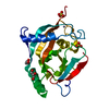 3kr7S C: citing same article ( S: Starting model for refinement |
|---|---|
| Similar structure data |


-Links
 PDBj
PDBj


- Assembly
| Deposited unit | 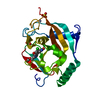
| ||||||||
|---|---|---|---|---|---|---|---|---|---|
| 1 |
| ||||||||
| Unit cell |
|
-Components
| #1: Protein | / TANK2 / ADP-ribosyltransferase diphtheria toxin-like 6 / ARTD6 / Poly [ADP-ribose] polymerase 5B / ...TANK2 / ADP-ribosyltransferase diphtheria toxin-like 6 / ARTD6 / Poly [ADP-ribose] polymerase 5B / TNKS-2 / TRF1-interacting ankyrin-related ADP-ribose polymerase 2 / Tankyrase II / Tankyrase-like protein / Tankyrase-related protein Mass: 23993.195 Da / Num. of mol.: 1 / Fragment: catalytic domain, UNP RESIDUES 952-1161 Source method: isolated from a genetically manipulated source Source: (gene. exp.) Homo sapiens (human) / Gene: PARP5B, TANK2, TNKL, TNKS2 / Plasmid: pNIC-Bsa4 / Production host:  Escherichia coli (E. coli) / Strain (production host): BL21 (DE3) / References: UniProt: Q9H2K2, NAD+ ADP-ribosyltransferase Escherichia coli (E. coli) / Strain (production host): BL21 (DE3) / References: UniProt: Q9H2K2, NAD+ ADP-ribosyltransferase | ||||
|---|---|---|---|---|---|
| #2: Chemical | ChemComp-ZN /   Mass: 65.409 Da / Num. of mol.: 1 / Source method: obtained synthetically / Formula: Zn Mass: 65.409 Da / Num. of mol.: 1 / Source method: obtained synthetically / Formula: Zn | ||||
| #3: Chemical | Sulfate  Mass: 96.063 Da / Num. of mol.: 2 / Source method: obtained synthetically / Formula: SO4 Mass: 96.063 Da / Num. of mol.: 2 / Source method: obtained synthetically / Formula: SO4#4: Chemical | ChemComp-AJ5 / |   Mass: 370.829 Da / Num. of mol.: 1 / Source method: obtained synthetically / Formula: C20H19ClN2O3 Mass: 370.829 Da / Num. of mol.: 1 / Source method: obtained synthetically / Formula: C20H19ClN2O3#5: Water | ChemComp-HOH / | Water Mass: 18.015 Da / Num. of mol.: 172 / Source method: isolated from a natural source / Formula: H2O Mass: 18.015 Da / Num. of mol.: 172 / Source method: isolated from a natural source / Formula: H2O |
-Experimental details
-Experiment
| Experiment | Method: X-RAY DIFFRACTION / Number of used crystals: 1 |
|---|
- Sample preparation
| Crystal | Density Matthews: 2.72 Å3/Da / Density % sol: 54.86 % |
|---|---|
| Crystal grow | Temperature: 277 K / Method: vapor diffusion, hanging drop / pH: 8.5 Details: 17% PEG 3350, 0.2M Ammonium sulphate, 0.1M Tris pH 8.5, vapour diffusion, hanging drop, temperature 277K |
-Data collection
| Diffraction | Mean temperature: 277 K |
|---|---|
| Diffraction source | Source: SYNCHROTRON / Site: Australian Synchrotron  / Beamline: MX1 / Beamline: MX1 |
| Detector | Type: ADSC QUANTUM 315r / Detector: CCD |
| Radiation | Protocol: SINGLE WAVELENGTH / Monochromatic (M) / Laue (L): M / Scattering type: x-ray |
| Radiation wavelength | Relative weight: 1 |
| Reflection | Resolution: 2.09→115.8 Å / Num. all: 16212 / Num. obs: 16212 / % possible obs: 98 % / Observed criterion σ(F): 0 / Observed criterion σ(I): 2 |
- Processing
| Software |
| |||||||||||||||||||||||||||||||||||||||||||||||||||||||||||||||||
|---|---|---|---|---|---|---|---|---|---|---|---|---|---|---|---|---|---|---|---|---|---|---|---|---|---|---|---|---|---|---|---|---|---|---|---|---|---|---|---|---|---|---|---|---|---|---|---|---|---|---|---|---|---|---|---|---|---|---|---|---|---|---|---|---|---|---|
| Refinement | Method to determine structure: MOLECULAR REPLACEMENT Starting model: 3KR7 Resolution: 2.09→67 Å / Cor.coef. Fo:Fc: 0.946 / Cor.coef. Fo:Fc free: 0.919 / Occupancy max: 1 / Occupancy min: 0.5 / SU B: 4.77 / SU ML: 0.126 / Cross valid method: THROUGHOUT / σ(F): 0 / ESU R: 0.201 / ESU R Free: 0.186 / Stereochemistry target values: MAXIMUM LIKELIHOOD Details: HYDROGENS HAVE BEEN ADDED IN THE RIDING POSITIONS U VALUES: REFINED INDIVIDUALLY
| |||||||||||||||||||||||||||||||||||||||||||||||||||||||||||||||||
| Solvent computation | Ion probe radii: 0.8 Å / Shrinkage radii: 0.8 Å / VDW probe radii: 1.4 Å / Solvent model: MASK | |||||||||||||||||||||||||||||||||||||||||||||||||||||||||||||||||
| Displacement parameters | Biso max: 105.48 Å2 / Biso mean: 27.9024 Å2 / Biso min: 10.4 Å2
| |||||||||||||||||||||||||||||||||||||||||||||||||||||||||||||||||
| Refinement step | Cycle: LAST / Resolution: 2.09→67 Å
| |||||||||||||||||||||||||||||||||||||||||||||||||||||||||||||||||
| Refine LS restraints |
| |||||||||||||||||||||||||||||||||||||||||||||||||||||||||||||||||
| LS refinement shell | Resolution: 2.09→2.144 Å / Total num. of bins used: 20
|
