 Movie
Movie Controller

[English] 日本語
 Yorodumi
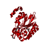
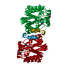
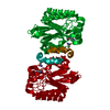
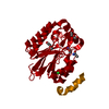
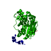
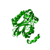
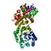
- PDB-4izw: The E41L mutant of the amidase from Nesterenkonia sp. AN1 showing... -
+ Open data

- Basic information
| Entry | Database: PDB / ID: 4izw | ||||||
|---|---|---|---|---|---|---|---|
| Title | The E41L mutant of the amidase from Nesterenkonia sp. AN1 showing covalent addition of the acetamide moiety of fluoroacetamide at the active site cysteine | ||||||
 Components Components | Amidase | ||||||
 Keywords Keywords | HYDROLASE / active site / fluoroacetamide / acetamide | ||||||
| Function / homology |  Function and homology informationamidase / indoleacetamide hydrolase activity / N-carbamoylputrescine amidase activity / beta-alanine biosynthetic process via 3-ureidopropionate / beta-ureidopropionase activity / putrescine biosynthetic process from arginine / amidase activity Function and homology informationamidase / indoleacetamide hydrolase activity / N-carbamoylputrescine amidase activity / beta-alanine biosynthetic process via 3-ureidopropionate / beta-ureidopropionase activity / putrescine biosynthetic process from arginine / amidase activitySimilarity search - Function | ||||||
| Biological species |  Nesterenkonia sp. 10004 (bacteria) Nesterenkonia sp. 10004 (bacteria) | ||||||
| Method | X-RAY DIFFRACTION / SYNCHROTRON / MOLECULAR REPLACEMENT / molecular replacement / Resolution: 1.6 Å | ||||||
 Authors Authors | Kimani, S.W. / Sewell, B.T. | ||||||
 Citation Citation | Journal: To be Published Title: Covalent modifications of the active site cysteine occur as a result of mutating the glutamate of the catalytic triad in the amidase from Nesterenkonia sp. Authors: Kimani, S.W. / Hunter, R. / Vlok, M. / Watermeyer, J. / Sewell, B.T. | ||||||
| History |
|
- Structure visualization
| Structure viewer | Molecule: MolmilJmol/JSmol |
|---|
- Downloads & links
-Download
| PDBx/mmCIF format | 4izw.cif.gz | 63.9 KB | Display | PDBx/mmCIF format |
|---|---|---|---|---|
| PDB format | pdb4izw.ent.gz | 49.6 KB | Display | PDB format |
| PDBx/mmJSON format | 4izw.json.gz | Tree view | PDBx/mmJSON format | |
| Others |  Other downloads Other downloads |
-Validation report
| Arichive directory | https://data.pdbj.org/pub/pdb/validation_reports/iz/4izwftp://data.pdbj.org/pub/pdb/validation_reports/iz/4izw | HTTPS FTP |
|---|
-Related structure data


-Links
 PDBj
PDBj


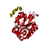
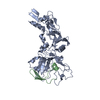
- Assembly
| Deposited unit | 
| |||||||||
|---|---|---|---|---|---|---|---|---|---|---|
| 1 | 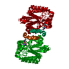
| |||||||||
| Unit cell |
| |||||||||
| Components on special symmetry positions |
|
-Components
| #1: Protein | Mass: 30084.795 Da / Num. of mol.: 1 / Mutation: E41Q Source method: isolated from a genetically manipulated source Source: (gene. exp.) Nesterenkonia sp. 10004 (bacteria) / Strain: AN1 / Gene: Nit2 / Plasmid: pET28a / Production host: Escherichia coli (E. coli) / Strain (production host): BL21(DE3) / References: UniProt: D0VWZ1, amidase | ||
|---|---|---|---|
| #2: Chemical | ChemComp-ACM / Acetamide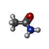  Mass: 59.067 Da / Num. of mol.: 1 / Source method: obtained synthetically / Formula: C2H5NO Mass: 59.067 Da / Num. of mol.: 1 / Source method: obtained synthetically / Formula: C2H5NO | ||
| #3: Chemical | Fluoroacetamide  Mass: 77.058 Da / Num. of mol.: 3 / Source method: obtained synthetically / Formula: C2H4FNO Mass: 77.058 Da / Num. of mol.: 3 / Source method: obtained synthetically / Formula: C2H4FNO#4: Water | ChemComp-HOH / | Water Mass: 18.015 Da / Num. of mol.: 162 / Source method: isolated from a natural source / Formula: H2O Mass: 18.015 Da / Num. of mol.: 162 / Source method: isolated from a natural source / Formula: H2O |
-Experimental details
-Experiment
| Experiment | Method: X-RAY DIFFRACTION / Number of used crystals: 1 |
|---|
- Sample preparation
| Crystal | Density Matthews: 2.33 Å3/Da / Density % sol: 47.16 % |
|---|---|
| Crystal grow | Temperature: 298 K / Method: vapor diffusion, sitting drop / pH: 7.5 Details: 0.1 M HEPES sodium, 2% PEG 400, 2.0 M ammonium sulfate, pH 7.5, VAPOR DIFFUSION, SITTING DROP, temperature 298K |
-Data collection
| Diffraction | Mean temperature: 100 K | ||||||||||||||||||||||||||||||||||||||||||||||||||||||||||||||||||||||||||||||||||||||||
|---|---|---|---|---|---|---|---|---|---|---|---|---|---|---|---|---|---|---|---|---|---|---|---|---|---|---|---|---|---|---|---|---|---|---|---|---|---|---|---|---|---|---|---|---|---|---|---|---|---|---|---|---|---|---|---|---|---|---|---|---|---|---|---|---|---|---|---|---|---|---|---|---|---|---|---|---|---|---|---|---|---|---|---|---|---|---|---|---|---|
| Diffraction source | Source: SYNCHROTRON / Site: ESRF  / Beamline: BM14 / Wavelength: 0.9184 Å / Beamline: BM14 / Wavelength: 0.9184 Å | ||||||||||||||||||||||||||||||||||||||||||||||||||||||||||||||||||||||||||||||||||||||||
| Detector | Detector: CCD / Date: Jul 10, 2010 | ||||||||||||||||||||||||||||||||||||||||||||||||||||||||||||||||||||||||||||||||||||||||
| Radiation | Protocol: SINGLE WAVELENGTH / Monochromatic (M) / Laue (L): M / Scattering type: x-ray | ||||||||||||||||||||||||||||||||||||||||||||||||||||||||||||||||||||||||||||||||||||||||
| Radiation wavelength | Wavelength: 0.9184 Å / Relative weight: 1 | ||||||||||||||||||||||||||||||||||||||||||||||||||||||||||||||||||||||||||||||||||||||||
| Reflection | Resolution: 1.6→42.87 Å / Num. obs: 35169 / % possible obs: 94 % / Redundancy: 2.61 % / Rmerge(I) obs: 0.076 / Χ2: 0.98 / Net I/σ(I): 7.9 / Scaling rejects: 695 | ||||||||||||||||||||||||||||||||||||||||||||||||||||||||||||||||||||||||||||||||||||||||
| Reflection shell | Diffraction-ID: 1
|
-Phasing
| Phasing | Method: molecular replacement | |||||||||
|---|---|---|---|---|---|---|---|---|---|---|
| Phasing MR | Rfactor: 31.3 / Model details: Phaser MODE: MR_AUTO
|
- Processing
| Software |
| |||||||||||||||||||||||||||||||||||||||||||||||||||||||||||||||||
|---|---|---|---|---|---|---|---|---|---|---|---|---|---|---|---|---|---|---|---|---|---|---|---|---|---|---|---|---|---|---|---|---|---|---|---|---|---|---|---|---|---|---|---|---|---|---|---|---|---|---|---|---|---|---|---|---|---|---|---|---|---|---|---|---|---|---|
| Refinement | Method to determine structure: MOLECULAR REPLACEMENT / Resolution: 1.6→42.87 Å / Cor.coef. Fo:Fc: 0.96 / Cor.coef. Fo:Fc free: 0.953 / WRfactor Rfree: 0.2186 / WRfactor Rwork: 0.1914 / Occupancy max: 1 / Occupancy min: 0.5 / FOM work R set: 0.8639 / SU B: 1.582 / SU ML: 0.054 / SU R Cruickshank DPI: 0.0902 / SU Rfree: 0.0903 / Cross valid method: THROUGHOUT / σ(F): 0 / ESU R: 0.09 / ESU R Free: 0.09 / Stereochemistry target values: MAXIMUM LIKELIHOOD Details: HYDROGENS HAVE BEEN ADDED IN THE RIDING POSITIONS U VALUES : REFINED INDIVIDUALLY
| |||||||||||||||||||||||||||||||||||||||||||||||||||||||||||||||||
| Solvent computation | Ion probe radii: 0.8 Å / Shrinkage radii: 0.8 Å / VDW probe radii: 1.4 Å / Solvent model: MASK | |||||||||||||||||||||||||||||||||||||||||||||||||||||||||||||||||
| Displacement parameters | Biso max: 72.62 Å2 / Biso mean: 19.9973 Å2 / Biso min: 9.85 Å2
| |||||||||||||||||||||||||||||||||||||||||||||||||||||||||||||||||
| Refinement step | Cycle: LAST / Resolution: 1.6→42.87 Å
| |||||||||||||||||||||||||||||||||||||||||||||||||||||||||||||||||
| Refine LS restraints |
| |||||||||||||||||||||||||||||||||||||||||||||||||||||||||||||||||
| LS refinement shell | Resolution: 1.6→1.642 Å / Total num. of bins used: 20
|
