 Movie
Movie Controller


+ Open data

- Basic information
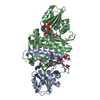
| Entry | Database: PDB / ID: 4iyp | ||||||
|---|---|---|---|---|---|---|---|
| Title | structure of the nPP2Ac-alpha4 complex | ||||||
 Components Components |
| ||||||
 Keywords Keywords |  SIGNALING PROTEIN / alpha4 / PP2A / latency / helix motif SIGNALING PROTEIN / alpha4 / PP2A / latency / helix motif | ||||||
| Function / homology |  Function and homology informationregulation of dephosphorylation / Integration of energy metabolism / PP2A-mediated dephosphorylation of key metabolic factors / regulation of microtubule-based movement / regulation of microtubule binding / MASTL Facilitates Mitotic Progression / protein phosphatase type 2A complex / peptidyl-serine dephosphorylation / peptidyl-threonine dephosphorylation / positive regulation of microtubule binding ...regulation of dephosphorylation / Integration of energy metabolism / PP2A-mediated dephosphorylation of key metabolic factors / regulation of microtubule-based movement / regulation of microtubule binding / MASTL Facilitates Mitotic Progression / protein phosphatase type 2A complex / peptidyl-serine dephosphorylation / peptidyl-threonine dephosphorylation / positive regulation of microtubule binding / Regulation of glycolysis by fructose 2,6-bisphosphate metabolism / Inhibition of replication initiation of damaged DNA by RB1/E2F1 / protein phosphatase regulator activity / GABA receptor binding / APC truncation mutants have impaired AXIN binding / AXIN missense mutants destabilize the destruction complex / Truncations of AMER1 destabilize the destruction complex / Initiation of Nuclear Envelope (NE) Reformation / ERKs are inactivated / negative regulation of epithelial to mesenchymal transition / Beta-catenin phosphorylation cascade / Signaling by GSK3beta mutants / CTNNB1 S33 mutants aren't phosphorylated / CTNNB1 S37 mutants aren't phosphorylated / CTNNB1 S45 mutants aren't phosphorylated / CTNNB1 T41 mutants aren't phosphorylated / negative regulation of stress-activated MAPK cascade / regulation of growth / Disassembly of the destruction complex and recruitment of AXIN to the membrane / negative regulation of glycolytic process through fructose-6-phosphate / positive regulation of NLRP3 inflammasome complex assembly / myosin phosphatase activity / CTLA4 inhibitory signaling / protein serine/threonine phosphatase activity / Platelet sensitization by LDL / B cell activation / protein-serine/threonine phosphatase / regulation of cell differentiation / ERK/MAPK targets / T cell homeostasis / regulation of G1/S transition of mitotic cell cycle / phosphoprotein phosphatase activity / mesoderm development / chromosome, centromeric region / DARPP-32 events / negative regulation of phosphatidylinositol 3-kinase/protein kinase B signal transduction / Nonsense Mediated Decay (NMD) enhanced by the Exon Junction Complex (EJC) / response to tumor necrosis factor / Amplification of signal from unattached kinetochores via a MAD2 inhibitory signal / Cyclin A/B1/B2 associated events during G2/M transition / Mitotic Prometaphase / EML4 and NUDC in mitotic spindle formation / Resolution of Sister Chromatid Cohesion / response to interleukin-1 / protein dephosphorylation / protein phosphatase 2A binding / meiotic cell cycle / protein tyrosine phosphatase activity / RHO GTPases Activate Formins / response to lead ion / regulation of protein phosphorylation / Spry regulation of FGF signaling / RAF activation / PKR-mediated signaling / Degradation of beta-catenin by the destruction complex / negative regulation of cysteine-type endopeptidase activity involved in apoptotic process / tau protein binding / positive regulation of protein serine/threonine kinase activity / spindle pole / Negative regulation of MAPK pathway / Separation of Sister Chromatids / Cyclin D associated events in G1 / microtubule cytoskeleton / Regulation of TP53 Degradation / mitotic cell cycle / PI5P, PP2A and IER3 Regulate PI3K/AKT Signaling / intracellular signal transduction / membrane raft / protein heterodimerization activity / synapse / negative regulation of transcription by RNA polymerase II / signal transduction / mitochondrion / extracellular exosome / membrane / metal ion binding / nucleus / plasma membrane / cytosol / cytoplasm Function and homology informationregulation of dephosphorylation / Integration of energy metabolism / PP2A-mediated dephosphorylation of key metabolic factors / regulation of microtubule-based movement / regulation of microtubule binding / MASTL Facilitates Mitotic Progression / protein phosphatase type 2A complex / peptidyl-serine dephosphorylation / peptidyl-threonine dephosphorylation / positive regulation of microtubule binding ...regulation of dephosphorylation / Integration of energy metabolism / PP2A-mediated dephosphorylation of key metabolic factors / regulation of microtubule-based movement / regulation of microtubule binding / MASTL Facilitates Mitotic Progression / protein phosphatase type 2A complex / peptidyl-serine dephosphorylation / peptidyl-threonine dephosphorylation / positive regulation of microtubule binding / Regulation of glycolysis by fructose 2,6-bisphosphate metabolism / Inhibition of replication initiation of damaged DNA by RB1/E2F1 / protein phosphatase regulator activity / GABA receptor binding / APC truncation mutants have impaired AXIN binding / AXIN missense mutants destabilize the destruction complex / Truncations of AMER1 destabilize the destruction complex / Initiation of Nuclear Envelope (NE) Reformation / ERKs are inactivated / negative regulation of epithelial to mesenchymal transition / Beta-catenin phosphorylation cascade / Signaling by GSK3beta mutants / CTNNB1 S33 mutants aren't phosphorylated / CTNNB1 S37 mutants aren't phosphorylated / CTNNB1 S45 mutants aren't phosphorylated / CTNNB1 T41 mutants aren't phosphorylated / negative regulation of stress-activated MAPK cascade / regulation of growth / Disassembly of the destruction complex and recruitment of AXIN to the membrane / negative regulation of glycolytic process through fructose-6-phosphate / positive regulation of NLRP3 inflammasome complex assembly / myosin phosphatase activity / CTLA4 inhibitory signaling / protein serine/threonine phosphatase activity / Platelet sensitization by LDL / B cell activation / protein-serine/threonine phosphatase / regulation of cell differentiation / ERK/MAPK targets / T cell homeostasis / regulation of G1/S transition of mitotic cell cycle / phosphoprotein phosphatase activity / mesoderm development / chromosome, centromeric region / DARPP-32 events / negative regulation of phosphatidylinositol 3-kinase/protein kinase B signal transduction / Nonsense Mediated Decay (NMD) enhanced by the Exon Junction Complex (EJC) / response to tumor necrosis factor / Amplification of signal from unattached kinetochores via a MAD2 inhibitory signal / Cyclin A/B1/B2 associated events during G2/M transition / Mitotic Prometaphase / EML4 and NUDC in mitotic spindle formation / Resolution of Sister Chromatid Cohesion / response to interleukin-1 / protein dephosphorylation / protein phosphatase 2A binding / meiotic cell cycle / protein tyrosine phosphatase activity / RHO GTPases Activate Formins / response to lead ion / regulation of protein phosphorylation / Spry regulation of FGF signaling / RAF activation / PKR-mediated signaling / Degradation of beta-catenin by the destruction complex / negative regulation of cysteine-type endopeptidase activity involved in apoptotic process / tau protein binding / positive regulation of protein serine/threonine kinase activity / spindle pole / Negative regulation of MAPK pathway / Separation of Sister Chromatids / Cyclin D associated events in G1 / microtubule cytoskeleton / Regulation of TP53 Degradation / mitotic cell cycle / PI5P, PP2A and IER3 Regulate PI3K/AKT Signaling / intracellular signal transduction / membrane raft / protein heterodimerization activity / synapse / negative regulation of transcription by RNA polymerase II / signal transduction / mitochondrion / extracellular exosome / membrane / metal ion binding / nucleus / plasma membrane / cytosol / cytoplasmSimilarity search - Function | ||||||
| Biological species |  Homo sapiens (human) Homo sapiens (human) | ||||||
| Method | X-RAY DIFFRACTION / SYNCHROTRON / SAD / Resolution: 2.797 Å | ||||||
 Authors Authors | Jiang, L. / Stanevich, V. / Satyshur, K.A. / Xing, Y. | ||||||
 Citation Citation | Journal: Nat Commun / Year: 2013 Title: Structural basis of protein phosphatase 2A stable latency. Authors: Jiang, L. / Stanevich, V. / Satyshur, K.A. / Kong, M. / Watkins, G.R. / Wadzinski, B.E. / Sengupta, R. / Xing, Y. | ||||||
| History |
|
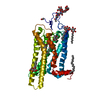
- Structure visualization
| Structure viewer | Molecule: MolmilJmol/JSmol |
|---|
- Downloads & links
-Download
| PDBx/mmCIF format | 4iyp.cif.gz | 147.8 KB | Display | PDBx/mmCIF format |
|---|---|---|---|---|
| PDB format | pdb4iyp.ent.gz | 123.4 KB | Display | PDB format |
| PDBx/mmJSON format | 4iyp.json.gz | Tree view | PDBx/mmJSON format | |
| Others |  Other downloads Other downloads |
-Validation report
| Arichive directory | https://data.pdbj.org/pub/pdb/validation_reports/iy/4iypftp://data.pdbj.org/pub/pdb/validation_reports/iy/4iyp | HTTPS FTP |
|---|
-Related structure data
| Related structure data | |
|---|---|
| Similar structure data |


-Links
 PDBj
PDBj


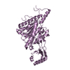
- Assembly
| Deposited unit | 
| ||||||||
|---|---|---|---|---|---|---|---|---|---|
| 1 |
| ||||||||
| Unit cell |
|
-Components
| #1: Protein | Mass: 25926.387 Da / Num. of mol.: 1 Source method: isolated from a genetically manipulated source Source: (gene. exp.) Homo sapiens (human) / Gene: alpha4, IBP1, IGBP1 / Plasmid: pQlink / Production host:  Escherichia coli (E. coli) / Strain (production host): DH5alpha / References: UniProt: P78318 Escherichia coli (E. coli) / Strain (production host): DH5alpha / References: UniProt: P78318 |
|---|---|
| #2: Protein | Mass: 17896.840 Da / Num. of mol.: 1 Source method: isolated from a genetically manipulated source Source: (gene. exp.) Homo sapiens (human) / Gene: PP2Ac, ppp2c, PPP2CA / Plasmid: pQlink / Production host: Escherichia coli (E. coli) / Strain (production host): PP2AReferences: UniProt: P67775, protein-serine/threonine phosphatase |
| #3: Water | ChemComp-HOH / Water Mass: 18.015 Da / Num. of mol.: 34 / Source method: isolated from a natural source / Formula: H2O Mass: 18.015 Da / Num. of mol.: 34 / Source method: isolated from a natural source / Formula: H2O |
-Experimental details
-Experiment
| Experiment | Method: X-RAY DIFFRACTION / Number of used crystals: 1 |
|---|
- Sample preparation
| Crystal | Density Matthews: 3.61 Å3/Da / Density % sol: 65.91 % |
|---|---|
| Crystal grow | Temperature: 291 K / Method: vapor diffusion, sitting drop / pH: 8 Details: 12-15% PEG3350 (v/v), 0.3 M Na/K tartrate, pH 8.0, VAPOR DIFFUSION, SITTING DROP, temperature 291K |
-Data collection
| Diffraction | Mean temperature: 100 K |
|---|---|
| Diffraction source | Source: SYNCHROTRON / Site: APS  / Beamline: 21-ID-F / Wavelength: 0.9792 Å / Beamline: 21-ID-F / Wavelength: 0.9792 Å |
| Detector | Type: MARMOSAIC 300 mm CCD / Detector: CCD / Date: Oct 18, 2010 |
| Radiation | Protocol: SINGLE WAVELENGTH / Monochromatic (M) / Laue (L): M / Scattering type: x-ray |
| Radiation wavelength | Wavelength: 0.9792 Å / Relative weight: 1 |
| Reflection | Resolution: 2.797→50 Å / Num. all: 15948 / Num. obs: 15948 / % possible obs: 99.8 % / Observed criterion σ(F): 0 / Observed criterion σ(I): 0 / Redundancy: 20.1 % / Rsym value: 0.07 / Net I/σ(I): 28.7 |
| Reflection shell | Resolution: 2.797→2.85 Å / Redundancy: 10.7 % / Mean I/σ(I) obs: 3.22 / Num. unique all: 777 / Rsym value: 0.545 / % possible all: 98.7 |
- Processing
| Software |
| |||||||||||||||||||||||||||||||||||||||||||||||||||||||||||||||||||||||||||||||||||||||||||||||||||||||||||||||||||||||||||||||||||||||||||||||||||||||||||||||||||||||||||||||||||||||||||||||||||||||||||||||||||||||||||||||||
|---|---|---|---|---|---|---|---|---|---|---|---|---|---|---|---|---|---|---|---|---|---|---|---|---|---|---|---|---|---|---|---|---|---|---|---|---|---|---|---|---|---|---|---|---|---|---|---|---|---|---|---|---|---|---|---|---|---|---|---|---|---|---|---|---|---|---|---|---|---|---|---|---|---|---|---|---|---|---|---|---|---|---|---|---|---|---|---|---|---|---|---|---|---|---|---|---|---|---|---|---|---|---|---|---|---|---|---|---|---|---|---|---|---|---|---|---|---|---|---|---|---|---|---|---|---|---|---|---|---|---|---|---|---|---|---|---|---|---|---|---|---|---|---|---|---|---|---|---|---|---|---|---|---|---|---|---|---|---|---|---|---|---|---|---|---|---|---|---|---|---|---|---|---|---|---|---|---|---|---|---|---|---|---|---|---|---|---|---|---|---|---|---|---|---|---|---|---|---|---|---|---|---|---|---|---|---|---|---|---|---|---|---|---|---|---|---|---|---|---|---|---|---|---|---|---|---|
| Refinement | Method to determine structure: SAD / Resolution: 2.797→34.428 Å / SU ML: 0.33 / σ(F): 0.61 / Phase error: 22.59 / Stereochemistry target values: ML
| |||||||||||||||||||||||||||||||||||||||||||||||||||||||||||||||||||||||||||||||||||||||||||||||||||||||||||||||||||||||||||||||||||||||||||||||||||||||||||||||||||||||||||||||||||||||||||||||||||||||||||||||||||||||||||||||||
| Solvent computation | Shrinkage radii: 0.9 Å / VDW probe radii: 1.11 Å / Solvent model: FLAT BULK SOLVENT MODEL | |||||||||||||||||||||||||||||||||||||||||||||||||||||||||||||||||||||||||||||||||||||||||||||||||||||||||||||||||||||||||||||||||||||||||||||||||||||||||||||||||||||||||||||||||||||||||||||||||||||||||||||||||||||||||||||||||
| Refinement step | Cycle: LAST / Resolution: 2.797→34.428 Å
| |||||||||||||||||||||||||||||||||||||||||||||||||||||||||||||||||||||||||||||||||||||||||||||||||||||||||||||||||||||||||||||||||||||||||||||||||||||||||||||||||||||||||||||||||||||||||||||||||||||||||||||||||||||||||||||||||
| Refine LS restraints |
| |||||||||||||||||||||||||||||||||||||||||||||||||||||||||||||||||||||||||||||||||||||||||||||||||||||||||||||||||||||||||||||||||||||||||||||||||||||||||||||||||||||||||||||||||||||||||||||||||||||||||||||||||||||||||||||||||
| LS refinement shell |
| |||||||||||||||||||||||||||||||||||||||||||||||||||||||||||||||||||||||||||||||||||||||||||||||||||||||||||||||||||||||||||||||||||||||||||||||||||||||||||||||||||||||||||||||||||||||||||||||||||||||||||||||||||||||||||||||||
| Refinement TLS params. | Method: refined / Refine-ID: X-RAY DIFFRACTION
| |||||||||||||||||||||||||||||||||||||||||||||||||||||||||||||||||||||||||||||||||||||||||||||||||||||||||||||||||||||||||||||||||||||||||||||||||||||||||||||||||||||||||||||||||||||||||||||||||||||||||||||||||||||||||||||||||
| Refinement TLS group |
|
