 Movie
Movie Controller

[English] 日本語
 Yorodumi
- PDB-4iyo: Crystal structure of cystathionine gamma lyase from Xanthomonas o... -
+ Open data

- Basic information
| Entry | Database: PDB / ID: 4iyo | |||||||||
|---|---|---|---|---|---|---|---|---|---|---|
| Title | Crystal structure of cystathionine gamma lyase from Xanthomonas oryzae pv. oryzae (XometC) in complex with E-site serine, A-site serine, A-site external aldimine structure with aminoacrylate and A-site iminopropionate intermediates | |||||||||
 Components Components | (Cystathionine gamma-lyase-like ... ) x 2 ) x 2 | |||||||||
 Keywords Keywords | LYASE / PLP DEPENDENT ENZYME / XOCGL / IMINOPROPIONATE / SERINE / Cys-Met metabolism PLP dependent enzyme / cystathionine gamma lyase / PLP binding | |||||||||
| Function / homology |  Function and homology information Function and homology informationcarbon-sulfur lyase activity / transsulfuration / pyridoxal phosphate bindingSimilarity search - Function | |||||||||
| Biological species |  Xanthomonas oryzae pv. oryzae (bacteria) Xanthomonas oryzae pv. oryzae (bacteria) | |||||||||
| Method | X-RAY DIFFRACTION / SYNCHROTRON / MOLECULAR REPLACEMENT / Resolution: 1.8 Å | |||||||||
 Authors Authors | Ngo, H.P.T. / Kim, J.K. / Kang, L.W. | |||||||||
 Citation Citation | Journal: Acta Crystallogr.,Sect.D / Year: 2014 Title: PLP undergoes conformational changes during the course of an enzymatic reaction. Authors: Ngo, H.P. / Cerqueira, N.M. / Kim, J.K. / Hong, M.K. / Fernandes, P.A. / Ramos, M.J. / Kang, L.W. | |||||||||
| History |
|
- Structure visualization
| Structure viewer | Molecule: MolmilJmol/JSmol |
|---|
- Downloads & links
-Download
| PDBx/mmCIF format | 4iyo.cif.gz | 324.9 KB | Display | PDBx/mmCIF format |
|---|---|---|---|---|
| PDB format | pdb4iyo.ent.gz | 260.8 KB | Display | PDB format |
| PDBx/mmJSON format | 4iyo.json.gz | Tree view | PDBx/mmJSON format | |
| Others |  Other downloads Other downloads |
-Validation report
| Arichive directory | https://data.pdbj.org/pub/pdb/validation_reports/iy/4iyoftp://data.pdbj.org/pub/pdb/validation_reports/iy/4iyo | HTTPS FTP |
|---|
-Related structure data
| Related structure data |  4ixsC  4ixzSC  4iy7C C: citing same article ( S: Starting model for refinement |
|---|---|
| Similar structure data |


-Links
 PDBj
PDBj

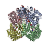
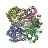
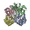
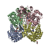
- Assembly
| Deposited unit | 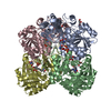
| ||||||||
|---|---|---|---|---|---|---|---|---|---|
| 1 |
| ||||||||
| Unit cell |
| ||||||||
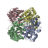
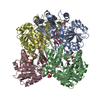
| Details | BIOMOLECULE: 1 SEE REMARK 350 FOR THE AUTHOR PROVIDED AND/OR PROGRAM GENERATED ASSEMBLY INFORMATION FOR THE STRUCTURE IN THIS ENTRY. THE REMARK MAY ALSO PROVIDE INFORMATION ON BURIED SURFACE AREA. COORDINATES FOR A COMPLETE MULTIMER REPRESENTING THE KNOWN BIOLOGICALLY SIGNIFICANT OLIGOMERIZATION STATE OF THE MOLECULE CAN BE GENERATED BY APPLYING BIOMT TRANSFORMATIONS GIVEN BELOW. BOTH NON-CRYSTALLOGRAPHIC AND CRYSTALLOGRAPHIC OPERATIONS ARE GIVEN. BIOMOLECULE: 1 AUTHOR DETERMINED BIOLOGICAL UNIT: TETRAMERIC SOFTWARE DETERMINED QUATERNARY STRUCTURE: TETRAMERIC SOFTWARE USED: PISA TOTAL BURIED SURFACE AREA: 21500 ANGSTROM**2 SURFACE AREA OF THE COMPLEX: 41940 ANGSTROM**2 CHANGE IN SOLVENT FREE ENERGY: -128.0 KCAL/MOL APPLY THE FOLLOWING TO CHAINS: A, B, C, D BIOMT1 1 1.000000 0.000000 0.000000 0.00000 BIOMT2 1 0.000000 1.000000 0.000000 0.00000 BIOMT3 1 0.000000 0.000000 1.000000 0.00000 |
-Components
-Cystathionine gamma-lyase-like ... , 2 types, 4 molecules ABCD
| #1: Protein | Mass: 42635.578 Da / Num. of mol.: 1 Source method: isolated from a genetically manipulated source Source: (gene. exp.) Xanthomonas oryzae pv. oryzae (bacteria)Strain: KACC 10331/KXO85 / Gene: metB, XOCGL, XOO0778 / Production host: Escherichia coli (E. coli) / References: UniProt: Q5H4T8, cystathionine gamma-lyase |
|---|---|
| #2: Protein | Mass: 42863.695 Da / Num. of mol.: 3 Source method: isolated from a genetically manipulated source Source: (gene. exp.) Xanthomonas oryzae pv. oryzae (bacteria)Strain: KACC 10331/KXO85 / Gene: metB, XOCGL, XOO0778 / Production host: Escherichia coli (E. coli) / References: UniProt: Q5H4T8, cystathionine gamma-lyase |
-Non-polymers , 7 types, 1337 molecules 


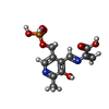


| #3: Chemical | ChemComp-SER / Serine Type: L-peptide linking / Mass: 105.093 Da / Num. of mol.: 5 / Source method: obtained synthetically / Formula: C3H7NO3 Type: L-peptide linking / Mass: 105.093 Da / Num. of mol.: 5 / Source method: obtained synthetically / Formula: C3H7NO3#4: Chemical | Glycerol Mass: 92.094 Da / Num. of mol.: 3 / Source method: obtained synthetically / Formula: C3H8O3 Mass: 92.094 Da / Num. of mol.: 3 / Source method: obtained synthetically / Formula: C3H8O3#5: Chemical | ChemComp-SO4 / | Sulfate Mass: 96.063 Da / Num. of mol.: 1 / Source method: obtained synthetically / Formula: SO4 Mass: 96.063 Da / Num. of mol.: 1 / Source method: obtained synthetically / Formula: SO4#6: Chemical | ChemComp-0JO / |  Mass: 316.204 Da / Num. of mol.: 1 / Source method: obtained synthetically / Formula: C11H13N2O7P Mass: 316.204 Da / Num. of mol.: 1 / Source method: obtained synthetically / Formula: C11H13N2O7P#7: Chemical |  Mass: 87.077 Da / Num. of mol.: 2 / Source method: obtained synthetically / Formula: C3H5NO2 Mass: 87.077 Da / Num. of mol.: 2 / Source method: obtained synthetically / Formula: C3H5NO2#8: Chemical | ChemComp-PYR / | Pyruvic acid Mass: 88.062 Da / Num. of mol.: 1 / Source method: obtained synthetically / Formula: C3H4O3 Mass: 88.062 Da / Num. of mol.: 1 / Source method: obtained synthetically / Formula: C3H4O3#9: Water | ChemComp-HOH / | WaterMass: 18.015 Da / Num. of mol.: 1324 / Source method: isolated from a natural source / Formula: H2O |
|---|
-Experimental details
-Experiment
| Experiment | Method: X-RAY DIFFRACTION / Number of used crystals: 1 |
|---|
- Sample preparation
| Crystal | Density Matthews: 2.17 Å3/Da / Density % sol: 43.44 % |
|---|---|
| Crystal grow | Temperature: 293 K / Method: vapor diffusion, hanging drop / pH: 9 Details: 25.5% PEG4000, 0.17M LITHIUM SULFATE, 0.085M TRIS, 15% GLYCEROL, PH 9.0, VAPOR DIFFUSION, HANGING DROP, TEMPERATURE 293K |
-Data collection
| Diffraction | Mean temperature: 100 K |
|---|---|
| Diffraction source | Source: SYNCHROTRON / Site: PAL/PLS  / Beamline: 4A / Wavelength: 1 / Wavelength: 1 Å / Beamline: 4A / Wavelength: 1 / Wavelength: 1 Å |
| Detector | Type: ADSC QUANTUM 210 / Detector: CCD / Date: Nov 8, 2010 |
| Radiation | Protocol: SINGLE WAVELENGTH / Monochromatic (M) / Laue (L): M / Scattering type: x-ray |
| Radiation wavelength | Wavelength: 1 Å / Relative weight: 1 |
| Reflection | Resolution: 1.78→50 Å / Num. obs: 142510 / % possible obs: 95.3 % / Observed criterion σ(F): 1 / Observed criterion σ(I): 1 |
| Reflection shell | Resolution: 1.78→1.81 Å / % possible all: 99.7 |
- Processing
| Software |
| |||||||||||||||||||||||||||||||||||||||||||||||||||||||||||||||||
|---|---|---|---|---|---|---|---|---|---|---|---|---|---|---|---|---|---|---|---|---|---|---|---|---|---|---|---|---|---|---|---|---|---|---|---|---|---|---|---|---|---|---|---|---|---|---|---|---|---|---|---|---|---|---|---|---|---|---|---|---|---|---|---|---|---|---|
| Refinement | Method to determine structure: MOLECULAR REPLACEMENT Starting model: PDB ENTRY 4IXZ Resolution: 1.8→33.82 Å / Cor.coef. Fo:Fc: 0.973 / Cor.coef. Fo:Fc free: 0.954 / SU B: 2.021 / SU ML: 0.064 / Cross valid method: THROUGHOUT / ESU R: 0.101 / ESU R Free: 0.104 / Stereochemistry target values: MAXIMUM LIKELIHOOD / Details: HYDROGENS HAVE BEEN ADDED IN THE RIDING POSITIONS
| |||||||||||||||||||||||||||||||||||||||||||||||||||||||||||||||||
| Solvent computation | Ion probe radii: 0.8 Å / Shrinkage radii: 0.8 Å / VDW probe radii: 1.4 Å / Solvent model: MASK | |||||||||||||||||||||||||||||||||||||||||||||||||||||||||||||||||
| Displacement parameters | Biso mean: 18.02 Å2
| |||||||||||||||||||||||||||||||||||||||||||||||||||||||||||||||||
| Refinement step | Cycle: LAST / Resolution: 1.8→33.82 Å
| |||||||||||||||||||||||||||||||||||||||||||||||||||||||||||||||||
| Refine LS restraints |
| |||||||||||||||||||||||||||||||||||||||||||||||||||||||||||||||||
| LS refinement shell | Resolution: 1.8→1.85 Å / Total num. of bins used: 20
|
