 Movie
Movie Controller


+ Open data

- Basic information
| Entry | Database: PDB / ID: 4iwb | ||||||
|---|---|---|---|---|---|---|---|
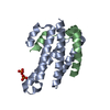
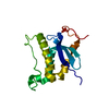
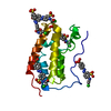
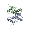
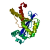
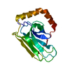
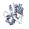
| Title | Novel Fold of FliC/FliS Fusion Protein | ||||||
 Components Components | FliC, FliS chimera | ||||||
 Keywords Keywords |  MOTOR PROTEIN / novel fold MOTOR PROTEIN / novel fold | ||||||
| Function / homology |  Function and homology information Function and homology informationbacterial-type flagellum / bacterial-type flagellum assembly / bacterial-type flagellum-dependent cell motility / structural molecule activity / extracellular region / cytosolSimilarity search - Function | ||||||
| Biological species |   Aquifex aeolicus (bacteria) Aquifex aeolicus (bacteria) | ||||||
| Method | X-RAY DIFFRACTION / SYNCHROTRON / MOLECULAR REPLACEMENT / Resolution: 1.75 Å | ||||||
 Authors Authors | Faham, S. | ||||||
 Citation Citation | Journal: Acta Crystallogr.,Sect.D / Year: 2013 Title: Protein design by fusion: implications for protein structure prediction and evolution. Authors: Skorupka, K. / Han, S.K. / Nam, H.J. / Kim, S. / Faham, S. | ||||||
| History |
|
- Structure visualization
| Structure viewer | Molecule: MolmilJmol/JSmol |
|---|
- Downloads & links
-Download
| PDBx/mmCIF format | 4iwb.cif.gz | 141.9 KB | Display | PDBx/mmCIF format |
|---|---|---|---|---|
| PDB format | pdb4iwb.ent.gz | 113.1 KB | Display | PDB format |
| PDBx/mmJSON format | 4iwb.json.gz | Tree view | PDBx/mmJSON format | |
| Others |  Other downloads Other downloads |
-Validation report
| Arichive directory | https://data.pdbj.org/pub/pdb/validation_reports/iw/4iwbftp://data.pdbj.org/pub/pdb/validation_reports/iw/4iwb | HTTPS FTP |
|---|
-Related structure data
| Related structure data | |
|---|---|
| Similar structure data |


-Links
 PDBj
PDBj- Assembly
| Deposited unit | 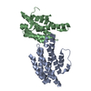
| ||||||||
|---|---|---|---|---|---|---|---|---|---|
| 1 | 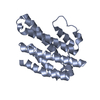
| ||||||||
| 2 | 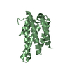
| ||||||||
| Unit cell |
|
-Components
| #1: Protein | Mass: 19292.281 Da / Num. of mol.: 2 / Fragment: SEE REMARK 999 Source method: isolated from a genetically manipulated source Source: (gene. exp.) Aquifex aeolicus (bacteria) / Gene: aq_1998, flaA, fliC, fliS / Plasmid: pBAD / Production host: Escherichia coli (E. coli) / Strain (production host): TOP10 / References: UniProt: O67803, UniProt: O67806#2: Water | ChemComp-HOH / | Water Mass: 18.015 Da / Num. of mol.: 161 / Source method: isolated from a natural source / Formula: H2O Mass: 18.015 Da / Num. of mol.: 161 / Source method: isolated from a natural source / Formula: H2OSequence details | PROTEIN IS A CHIMERA OF FLAGELLAR PROTEINS FLIC (UNP RESIDUES 478-518) AND FLIS (UNP RESIDUES 5-124) ...PROTEIN IS A CHIMERA OF FLAGELLAR PROTEINS FLIC (UNP RESIDUES 478-518) AND FLIS (UNP RESIDUES 5-124) CONNECTED BY A GLYCINE LINKER. | |
|---|
-Experimental details
-Experiment
| Experiment | Method: X-RAY DIFFRACTION / Number of used crystals: 1 |
|---|
- Sample preparation
| Crystal | Density Matthews: 2.55 Å3/Da / Density % sol: 51.69 % |
|---|---|
| Crystal grow | Temperature: 290 K / Method: vapor diffusion, hanging drop / pH: 9 Details: 38% PEG500 MME, 0.1 M sodium chloride, 0.1 M Tris-HCl, pH 9.0, VAPOR DIFFUSION, HANGING DROP, temperature 290K |
-Data collection
| Diffraction | Mean temperature: 103 K |
|---|---|
| Diffraction source | Source: SYNCHROTRON / Site: APS  / Beamline: 22-ID / Wavelength: 1 Å / Beamline: 22-ID / Wavelength: 1 Å |
| Detector | Type: MARMOSAIC 300 mm CCD / Detector: CCD |
| Radiation | Monochromator: Rosenbaum-Rock double-crystal Si(111) / Protocol: SINGLE WAVELENGTH / Monochromatic (M) / Laue (L): M / Scattering type: x-ray |
| Radiation wavelength | Wavelength: 1 Å / Relative weight: 1 |
| Reflection | Resolution: 1.75→50 Å / Num. all: 39366 / Num. obs: 39366 / % possible obs: 96.8 % / Observed criterion σ(F): 0 / Observed criterion σ(I): 0 / Redundancy: 5.9 % / Rmerge(I) obs: 0.073 / Net I/σ(I): 19.7 |
| Reflection shell | Resolution: 1.75→1.79 Å / Redundancy: 4 % / Rmerge(I) obs: 0.441 / Num. unique all: 2344 / % possible all: 88.2 |
- Processing
| Software |
| ||||||||||||||||||||||||||||||||||||||||||||||||||||||||||||||||||||||||||||||||||||||||||||||||||||||||||||||||||||||||||||||||||||||||||||||||||||||||||||||||||||||||||
|---|---|---|---|---|---|---|---|---|---|---|---|---|---|---|---|---|---|---|---|---|---|---|---|---|---|---|---|---|---|---|---|---|---|---|---|---|---|---|---|---|---|---|---|---|---|---|---|---|---|---|---|---|---|---|---|---|---|---|---|---|---|---|---|---|---|---|---|---|---|---|---|---|---|---|---|---|---|---|---|---|---|---|---|---|---|---|---|---|---|---|---|---|---|---|---|---|---|---|---|---|---|---|---|---|---|---|---|---|---|---|---|---|---|---|---|---|---|---|---|---|---|---|---|---|---|---|---|---|---|---|---|---|---|---|---|---|---|---|---|---|---|---|---|---|---|---|---|---|---|---|---|---|---|---|---|---|---|---|---|---|---|---|---|---|---|---|---|---|---|---|---|
| Refinement | Method to determine structure: MOLECULAR REPLACEMENT / Resolution: 1.75→29.09 Å / Cor.coef. Fo:Fc: 0.944 / Cor.coef. Fo:Fc free: 0.933 / SU B: 4.354 / SU ML: 0.065 / Cross valid method: THROUGHOUT / ESU R: 0.124 / ESU R Free: 0.115 / Stereochemistry target values: MAXIMUM LIKELIHOOD
| ||||||||||||||||||||||||||||||||||||||||||||||||||||||||||||||||||||||||||||||||||||||||||||||||||||||||||||||||||||||||||||||||||||||||||||||||||||||||||||||||||||||||||
| Solvent computation | Ion probe radii: 0.8 Å / Shrinkage radii: 0.8 Å / VDW probe radii: 1.4 Å / Solvent model: MASK | ||||||||||||||||||||||||||||||||||||||||||||||||||||||||||||||||||||||||||||||||||||||||||||||||||||||||||||||||||||||||||||||||||||||||||||||||||||||||||||||||||||||||||
| Displacement parameters | Biso mean: 30.27 Å2
| ||||||||||||||||||||||||||||||||||||||||||||||||||||||||||||||||||||||||||||||||||||||||||||||||||||||||||||||||||||||||||||||||||||||||||||||||||||||||||||||||||||||||||
| Refinement step | Cycle: LAST / Resolution: 1.75→29.09 Å
| ||||||||||||||||||||||||||||||||||||||||||||||||||||||||||||||||||||||||||||||||||||||||||||||||||||||||||||||||||||||||||||||||||||||||||||||||||||||||||||||||||||||||||
| Refine LS restraints |
| ||||||||||||||||||||||||||||||||||||||||||||||||||||||||||||||||||||||||||||||||||||||||||||||||||||||||||||||||||||||||||||||||||||||||||||||||||||||||||||||||||||||||||
| LS refinement shell | Resolution: 1.75→1.793 Å / Total num. of bins used: 20
| ||||||||||||||||||||||||||||||||||||||||||||||||||||||||||||||||||||||||||||||||||||||||||||||||||||||||||||||||||||||||||||||||||||||||||||||||||||||||||||||||||||||||||
| Refinement TLS params. | Method: refined / Refine-ID: X-RAY DIFFRACTION
| ||||||||||||||||||||||||||||||||||||||||||||||||||||||||||||||||||||||||||||||||||||||||||||||||||||||||||||||||||||||||||||||||||||||||||||||||||||||||||||||||||||||||||
| Refinement TLS group |
|
