 Movie
Movie Controller

[English] 日本語
 Yorodumi
- PDB-4irx: Crystal structure of Caulobacter myo-inositol binding protein bou... -
+ Open data

- Basic information
| Entry | Database: PDB / ID: 4irx | ||||||
|---|---|---|---|---|---|---|---|
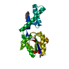
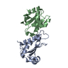
| Title | Crystal structure of Caulobacter myo-inositol binding protein bound to myo-inositol | ||||||
 Components Components | Sugar ABC transporter, periplasmic sugar-binding protein | ||||||
 Keywords Keywords |  TRANSPORT PROTEIN / ABC transporter / periplasmic binding protein / nutrient uptake / myo-inositol / selenomethionine TRANSPORT PROTEIN / ABC transporter / periplasmic binding protein / nutrient uptake / myo-inositol / selenomethionine | ||||||
| Function / homology |  Function and homology information Function and homology information | ||||||
| Biological species |  Caulobacter crescentus (bacteria) Caulobacter crescentus (bacteria) | ||||||
| Method | X-RAY DIFFRACTION / SYNCHROTRON / SAD / Resolution: 1.451 Å | ||||||
 Authors Authors | Herrou, J. / Crosson, S. | ||||||
 Citation Citation | Journal: J.Bacteriol. / Year: 2013 Title: myo-inositol and D-ribose ligand discrimination in an ABC periplasmic binding protein. Authors: Herrou, J. / Crosson, S. | ||||||
| History |
|
- Structure visualization
| Structure viewer | Molecule: MolmilJmol/JSmol |
|---|
- Downloads & links
-Download
| PDBx/mmCIF format | 4irx.cif.gz | 130.4 KB | Display | PDBx/mmCIF format |
|---|---|---|---|---|
| PDB format | pdb4irx.ent.gz | 107.8 KB | Display | PDB format |
| PDBx/mmJSON format | 4irx.json.gz | Tree view | PDBx/mmJSON format | |
| Others |  Other downloads Other downloads |
-Validation report
| Arichive directory | https://data.pdbj.org/pub/pdb/validation_reports/ir/4irxftp://data.pdbj.org/pub/pdb/validation_reports/ir/4irx | HTTPS FTP |
|---|
-Related structure data
| Similar structure data |
|---|


-Links
 PDBj
PDBj

- Assembly
| Deposited unit | 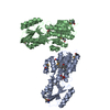
| ||||||||||||
|---|---|---|---|---|---|---|---|---|---|---|---|---|---|
| 1 | 
| ||||||||||||
| 2 | 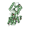
| ||||||||||||
| Unit cell |
| ||||||||||||
| Components on special symmetry positions |
|
-Components
| #1: Protein | Mass: 31116.289 Da / Num. of mol.: 2 / Fragment: UNP residues 39-325 Source method: isolated from a genetically manipulated source Source: (gene. exp.) Caulobacter crescentus (bacteria) / Strain: NA1000 / Gene: CC_0859 / Plasmid: pET28c / Production host: Escherichia coli (E. coli) / Strain (production host): Rosetta (DE3) pLysS / References: UniProt: Q9A9V2, UniProt: A0A0H3C834*PLUS#2: Chemical | Inositol  Mass: 180.156 Da / Num. of mol.: 2 / Source method: obtained synthetically / Formula: C6H12O6 / Comment: neurotransmitter, hormone*YM Mass: 180.156 Da / Num. of mol.: 2 / Source method: obtained synthetically / Formula: C6H12O6 / Comment: neurotransmitter, hormone*YM#3: Water | ChemComp-HOH / | Water Mass: 18.015 Da / Num. of mol.: 614 / Source method: isolated from a natural source / Formula: H2O Mass: 18.015 Da / Num. of mol.: 614 / Source method: isolated from a natural source / Formula: H2O |
|---|
-Experimental details
-Experiment
| Experiment | Method: X-RAY DIFFRACTION / Number of used crystals: 1 |
|---|
- Sample preparation
| Crystal | Density Matthews: 2.07 Å3/Da / Density % sol: 40.62 % |
|---|---|
| Crystal grow | Temperature: 292 K / Method: vapor diffusion, hanging drop / pH: 7.4 Details: 75 mN HEPES pH7.4, PEG3350 25%, 5 mM myo-inositol, VAPOR DIFFUSION, HANGING DROP, temperature 292K |
-Data collection
| Diffraction | Mean temperature: 100 K |
|---|---|
| Diffraction source | Source: SYNCHROTRON / Site: APS  / Beamline: 21-ID-D / Wavelength: 0.9794 Å / Beamline: 21-ID-D / Wavelength: 0.9794 Å |
| Detector | Type: MARMOSAIC 300 mm CCD / Detector: CCD / Date: Nov 27, 2011 / Details: mirrors |
| Radiation | Monochromator: Si 111 CHANNEL / Protocol: SINGLE WAVELENGTH / Monochromatic (M) / Laue (L): M / Scattering type: x-ray |
| Radiation wavelength | Wavelength: 0.9794 Å / Relative weight: 1 |
| Reflection | Resolution: 1.45→19.9 Å / Num. all: 78215 / Num. obs: 77441 / % possible obs: 99 % / Observed criterion σ(F): 2 / Observed criterion σ(I): -3 / Redundancy: 4.2 % / Biso Wilson estimate: 16.9 Å2 / Rmerge(I) obs: 0.08 / Net I/σ(I): 14.8 |
- Processing
| Software |
| |||||||||||||||||||||||||||||||||||||||||||||||||||||||||||||||||||||||||||||||||||||||||||||||||||||||||
|---|---|---|---|---|---|---|---|---|---|---|---|---|---|---|---|---|---|---|---|---|---|---|---|---|---|---|---|---|---|---|---|---|---|---|---|---|---|---|---|---|---|---|---|---|---|---|---|---|---|---|---|---|---|---|---|---|---|---|---|---|---|---|---|---|---|---|---|---|---|---|---|---|---|---|---|---|---|---|---|---|---|---|---|---|---|---|---|---|---|---|---|---|---|---|---|---|---|---|---|---|---|---|---|---|---|---|
| Refinement | Method to determine structure: SAD / Resolution: 1.451→19.9 Å / SU ML: 0.13 / σ(F): 0.2 / Phase error: 19.09 / Stereochemistry target values: ML
| |||||||||||||||||||||||||||||||||||||||||||||||||||||||||||||||||||||||||||||||||||||||||||||||||||||||||
| Solvent computation | Shrinkage radii: 0.9 Å / VDW probe radii: 1.11 Å / Solvent model: FLAT BULK SOLVENT MODEL | |||||||||||||||||||||||||||||||||||||||||||||||||||||||||||||||||||||||||||||||||||||||||||||||||||||||||
| Refinement step | Cycle: LAST / Resolution: 1.451→19.9 Å
| |||||||||||||||||||||||||||||||||||||||||||||||||||||||||||||||||||||||||||||||||||||||||||||||||||||||||
| Refine LS restraints |
| |||||||||||||||||||||||||||||||||||||||||||||||||||||||||||||||||||||||||||||||||||||||||||||||||||||||||
| LS refinement shell |
|
