 Movie
Movie Controller

[English] 日本語
 Yorodumi
- PDB-4inn: Protein HP1028 from the human pathogen Helicobacter pylori belong... -
+ Open data

- Basic information
| Entry | Database: PDB / ID: 4inn | ||||||
|---|---|---|---|---|---|---|---|
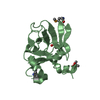
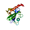
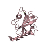
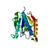
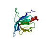
| Title | Protein HP1028 from the human pathogen Helicobacter pylori belongs to the lipocalin family | ||||||
 Components Components | HP1028 | ||||||
 Keywords Keywords |  TRANSPORT PROTEIN / beta-barrel / lipocalins fold / binding protein TRANSPORT PROTEIN / beta-barrel / lipocalins fold / binding protein | ||||||
| Function / homology | Lipocalin - #520 / Domain of unknown function DUF2147 / Uncharacterized protein conserved in bacteria (DUF2147) / Lipocalin / Beta Barrel / Mainly Beta / DUF2147 domain-containing protein Function and homology information Function and homology information | ||||||
| Biological species |   Helicobacter pylori (bacteria) Helicobacter pylori (bacteria) | ||||||
| Method | X-RAY DIFFRACTION / SYNCHROTRON / SAD / Resolution: 2.6 Å | ||||||
 Authors Authors | Barison, N. / Cendron, L. / Loconte, V. / Zanotti, G. | ||||||
 Citation Citation | Journal: Acta Crystallogr.,Sect.D / Year: 2013 Title: Protein HP1028 from the human pathogen Helicobacter pylori belongs to the lipocalin family. Authors: Barison, N. / Cendron, L. / Loconte, V. / Proctor, E.A. / Dokholyan, N.V. / Zanotti, G. | ||||||
| History |
|
- Structure visualization
| Structure viewer | Molecule: MolmilJmol/JSmol |
|---|
- Downloads & links
-Download
| PDBx/mmCIF format | 4inn.cif.gz | 71.7 KB | Display | PDBx/mmCIF format |
|---|---|---|---|---|
| PDB format | pdb4inn.ent.gz | 57.6 KB | Display | PDB format |
| PDBx/mmJSON format | 4inn.json.gz | Tree view | PDBx/mmJSON format | |
| Others |  Other downloads Other downloads |
-Validation report
| Arichive directory | https://data.pdbj.org/pub/pdb/validation_reports/in/4innftp://data.pdbj.org/pub/pdb/validation_reports/in/4inn | HTTPS FTP |
|---|
-Related structure data
| Similar structure data |
|---|


-Links
 PDBj
PDBj- Assembly
| Deposited unit | 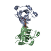
| |||||||||||||||||||||||||||||||||||||||
|---|---|---|---|---|---|---|---|---|---|---|---|---|---|---|---|---|---|---|---|---|---|---|---|---|---|---|---|---|---|---|---|---|---|---|---|---|---|---|---|---|
| 1 | 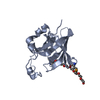
| |||||||||||||||||||||||||||||||||||||||
| 2 | 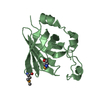
| |||||||||||||||||||||||||||||||||||||||
| Unit cell |
| |||||||||||||||||||||||||||||||||||||||
| Noncrystallographic symmetry (NCS) | NCS domain:
NCS domain segments:
NCS oper:
|
-Components
| #1: Protein | Mass: 20880.230 Da / Num. of mol.: 2 / Fragment: UNP residues 17-161 / Mutation: L129M Source method: isolated from a genetically manipulated source Source: (gene. exp.) Helicobacter pylori (bacteria) / Strain: CCUG17874 / Gene: hp1028, HP17_02362 / Plasmid: pET151-hp1028 / Production host: Escherichia coli (E. coli) / Strain (production host): BL21(DE3) / References: UniProt: I0ZEL6#2: Chemical | ChemComp-P33 / | Polyethylene glycol  Mass: 326.383 Da / Num. of mol.: 1 / Source method: obtained synthetically / Formula: C14H30O8 / Comment: precipitant*YM Mass: 326.383 Da / Num. of mol.: 1 / Source method: obtained synthetically / Formula: C14H30O8 / Comment: precipitant*YM#3: Water | ChemComp-HOH / | Water Mass: 18.015 Da / Num. of mol.: 64 / Source method: isolated from a natural source / Formula: H2O Mass: 18.015 Da / Num. of mol.: 64 / Source method: isolated from a natural source / Formula: H2O |
|---|
-Experimental details
-Experiment
| Experiment | Method: X-RAY DIFFRACTION / Number of used crystals: 1 |
|---|
- Sample preparation
| Crystal | Density Matthews: 2.14 Å3/Da / Density % sol: 42.42 % |
|---|---|
| Crystal grow | Temperature: 298 K / Method: vapor diffusion / pH: 8.5 Details: 0.2 M lithium sulfate, 0.1 M Tris-HCl, 30% PEG4000, pH 8.5, VAPOR DIFFUSION, temperature 298K |
-Data collection
| Diffraction | Mean temperature: 100 K |
|---|---|
| Diffraction source | Source: SYNCHROTRON / Site: ESRF  / Beamline: ID14-4 / Wavelength: 0.9795 Å / Beamline: ID14-4 / Wavelength: 0.9795 Å |
| Detector | Type: ADSC QUANTUM 315r / Detector: CCD / Date: Nov 21, 2008 |
| Radiation | Monochromator: channel cut Si(111) / Protocol: SINGLE WAVELENGTH / Monochromatic (M) / Laue (L): M / Scattering type: x-ray |
| Radiation wavelength | Wavelength: 0.9795 Å / Relative weight: 1 |
| Reflection | Resolution: 2.6→50.71 Å / Num. all: 11665 / Num. obs: 11665 / % possible obs: 100 % / Observed criterion σ(F): 0 / Observed criterion σ(I): 0 / Redundancy: 13.4 % / Rmerge(I) obs: 0.108 / Net I/σ(I): 19 |
| Reflection shell | Resolution: 2.6→2.74 Å / Redundancy: 13.9 % / Rmerge(I) obs: 0.225 / Mean I/σ(I) obs: 9.5 / Num. unique all: 1656 / % possible all: 99.9 |
- Processing
| Software |
| ||||||||||||||||||||||||||||||||||||||||||||||||||||||||||||||||||||||||||||||||||||||||||||||||||||||||||||||||||||||||||||||||||||||||||||||||||||||||||||||||||||||||||
|---|---|---|---|---|---|---|---|---|---|---|---|---|---|---|---|---|---|---|---|---|---|---|---|---|---|---|---|---|---|---|---|---|---|---|---|---|---|---|---|---|---|---|---|---|---|---|---|---|---|---|---|---|---|---|---|---|---|---|---|---|---|---|---|---|---|---|---|---|---|---|---|---|---|---|---|---|---|---|---|---|---|---|---|---|---|---|---|---|---|---|---|---|---|---|---|---|---|---|---|---|---|---|---|---|---|---|---|---|---|---|---|---|---|---|---|---|---|---|---|---|---|---|---|---|---|---|---|---|---|---|---|---|---|---|---|---|---|---|---|---|---|---|---|---|---|---|---|---|---|---|---|---|---|---|---|---|---|---|---|---|---|---|---|---|---|---|---|---|---|---|---|
| Refinement | Method to determine structure: SAD / Resolution: 2.6→50.67 Å / Cor.coef. Fo:Fc: 0.933 / Cor.coef. Fo:Fc free: 0.894 / SU B: 10.303 / SU ML: 0.226 / Cross valid method: THROUGHOUT / ESU R: 0.784 / ESU R Free: 0.312 / Stereochemistry target values: MAXIMUM LIKELIHOOD Details: HYDROGENS HAVE BEEN ADDED IN THE RIDING POSITIONS U VALUES : REFINED INDIVIDUALLY
| ||||||||||||||||||||||||||||||||||||||||||||||||||||||||||||||||||||||||||||||||||||||||||||||||||||||||||||||||||||||||||||||||||||||||||||||||||||||||||||||||||||||||||
| Solvent computation | Ion probe radii: 0.8 Å / Shrinkage radii: 0.8 Å / VDW probe radii: 1.2 Å / Solvent model: BABINET MODEL WITH MASK | ||||||||||||||||||||||||||||||||||||||||||||||||||||||||||||||||||||||||||||||||||||||||||||||||||||||||||||||||||||||||||||||||||||||||||||||||||||||||||||||||||||||||||
| Displacement parameters | Biso mean: 26.95 Å2
| ||||||||||||||||||||||||||||||||||||||||||||||||||||||||||||||||||||||||||||||||||||||||||||||||||||||||||||||||||||||||||||||||||||||||||||||||||||||||||||||||||||||||||
| Refinement step | Cycle: LAST / Resolution: 2.6→50.67 Å
| ||||||||||||||||||||||||||||||||||||||||||||||||||||||||||||||||||||||||||||||||||||||||||||||||||||||||||||||||||||||||||||||||||||||||||||||||||||||||||||||||||||||||||
| Refine LS restraints |
| ||||||||||||||||||||||||||||||||||||||||||||||||||||||||||||||||||||||||||||||||||||||||||||||||||||||||||||||||||||||||||||||||||||||||||||||||||||||||||||||||||||||||||
| Refine LS restraints NCS | Dom-ID: 1 / Auth asym-ID: A / Ens-ID: 1 / Refine-ID: X-RAY DIFFRACTION
| ||||||||||||||||||||||||||||||||||||||||||||||||||||||||||||||||||||||||||||||||||||||||||||||||||||||||||||||||||||||||||||||||||||||||||||||||||||||||||||||||||||||||||
| LS refinement shell | Resolution: 2.6→2.668 Å / Total num. of bins used: 20
|
