 Movie
Movie Controller

[English] 日本語
 Yorodumi
- PDB-4ina: Crystal Structure of the Q7MSS8_WOLSU protein from Wolinella succ... -
+ Open data

- Basic information
| Entry | Database: PDB / ID: 4ina | ||||||
|---|---|---|---|---|---|---|---|
| Title | Crystal Structure of the Q7MSS8_WOLSU protein from Wolinella succinogenes. Northeast Structural Genomics Consortium Target WsR35 | ||||||
 Components Components | saccharopine dehydrogenase | ||||||
 Keywords Keywords | OXIDOREDUCTASE / Structural Genomics / PSI-Biology / Northeast Structural Genomics Consortium / NESG / Saccharopine Dehydrogenase | ||||||
| Function / homology |  Function and homology information Function and homology information | ||||||
| Biological species |  Wolinella succinogenes (bacteria) Wolinella succinogenes (bacteria) | ||||||
| Method | X-RAY DIFFRACTION / SYNCHROTRON / SAD / Resolution: 2.488 Å | ||||||
 Authors Authors | Vorobiev, S. / Su, M. / Seetharaman, J. / Maglaqui, M. / Xiao, R. / Kohan, E. / Wang, D. / Everett, J.K. / Acton, T.B. / Montelione, G.T. ...Vorobiev, S. / Su, M. / Seetharaman, J. / Maglaqui, M. / Xiao, R. / Kohan, E. / Wang, D. / Everett, J.K. / Acton, T.B. / Montelione, G.T. / Tong, L. / Hunt, J.F. / Northeast Structural Genomics Consortium (NESG) | ||||||
 Citation Citation | Journal: To be Published Title: Crystal Structure of the Q7MSS8_WOLSU protein from Wolinella succinogenes. Authors: Vorobiev, S. / Su, M. / Seetharaman, J. / Maglaqui, M. / Xiao, R. / Kohan, E. / Wang, D. / Everett, J.K. / Acton, T.B. / Montelione, G.T. / Tong, L. / Hunt, J.F. | ||||||
| History |
|
- Structure visualization
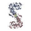
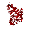
| Structure viewer | Molecule: MolmilJmol/JSmol |
|---|
- Downloads & links
-Download
| PDBx/mmCIF format | 4ina.cif.gz | 313.9 KB | Display | PDBx/mmCIF format |
|---|---|---|---|---|
| PDB format | pdb4ina.ent.gz | 267.5 KB | Display | PDB format |
| PDBx/mmJSON format | 4ina.json.gz | Tree view | PDBx/mmJSON format | |
| Others |  Other downloads Other downloads |
-Validation report
| Arichive directory | https://data.pdbj.org/pub/pdb/validation_reports/in/4inaftp://data.pdbj.org/pub/pdb/validation_reports/in/4ina | HTTPS FTP |
|---|
-Related structure data
| Similar structure data | |
|---|---|
| Other databases |


-Links
 PDBj
PDBj- Assembly
| Deposited unit | 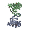
| ||||||||
|---|---|---|---|---|---|---|---|---|---|
| 1 |
| ||||||||
| Unit cell |
|
-Components
| #1: Protein | Mass: 46660.863 Da / Num. of mol.: 2 Source method: isolated from a genetically manipulated source Source: (gene. exp.) Wolinella succinogenes (bacteria) / Gene: WS0167 / Plasmid: WsR35-21.2 / Production host: Escherichia coli (E. coli) / Strain (production host): BL21(DE3)+ MagicReferences: UniProt: Q7MSS8, saccharopine dehydrogenase (NAD+, L-lysine-forming)#2: Water | ChemComp-HOH / | Water Mass: 18.015 Da / Num. of mol.: 146 / Source method: isolated from a natural source / Formula: H2O Mass: 18.015 Da / Num. of mol.: 146 / Source method: isolated from a natural source / Formula: H2O |
|---|
-Experimental details
-Experiment
| Experiment | Method: X-RAY DIFFRACTION / Number of used crystals: 1 |
|---|
- Sample preparation
| Crystal | Density Matthews: 2.33 Å3/Da / Density % sol: 47.31 % |
|---|---|
| Crystal grow | Temperature: 277 K / Method: microbatch crystallization under oil / pH: 6.5 Details: 20% PEG MME 5000, 0.1M BIS-TRIS, pH 6.5, Microbatch crystallization under oil, temperature 277K |
-Data collection
| Diffraction | Mean temperature: 100 K |
|---|---|
| Diffraction source | Source: SYNCHROTRON / Site: APS  / Beamline: 24-ID-E / Wavelength: 0.97923 Å / Beamline: 24-ID-E / Wavelength: 0.97923 Å |
| Detector | Type: ADSC QUANTUM 315 / Detector: CCD / Date: Dec 19, 2012 |
| Radiation | Protocol: SINGLE WAVELENGTH / Monochromatic (M) / Laue (L): M / Scattering type: x-ray |
| Radiation wavelength | Wavelength: 0.97923 Å / Relative weight: 1 |
| Reflection | Resolution: 2.49→50 Å / Num. all: 59291 / Num. obs: 57476 / % possible obs: 96.9 % / Observed criterion σ(F): 0 / Observed criterion σ(I): 0 / Redundancy: 3.7 % / Biso Wilson estimate: 35.49 Å2 / Rmerge(I) obs: 0.087 / Net I/σ(I): 10.6 |
| Reflection shell | Resolution: 2.49→2.6 Å / Redundancy: 3.7 % / Rmerge(I) obs: 0.474 / Mean I/σ(I) obs: 2.6 / Num. unique all: 7303 / % possible all: 97.7 |
- Processing
| Software |
| ||||||||||||||||||||||||||||||||||||||||||||||||||||||||||||||||||||||||||||||||||||||||||||||||||||||||||||||||||||||||||||||||||||||||||||||||||||||||||
|---|---|---|---|---|---|---|---|---|---|---|---|---|---|---|---|---|---|---|---|---|---|---|---|---|---|---|---|---|---|---|---|---|---|---|---|---|---|---|---|---|---|---|---|---|---|---|---|---|---|---|---|---|---|---|---|---|---|---|---|---|---|---|---|---|---|---|---|---|---|---|---|---|---|---|---|---|---|---|---|---|---|---|---|---|---|---|---|---|---|---|---|---|---|---|---|---|---|---|---|---|---|---|---|---|---|---|---|---|---|---|---|---|---|---|---|---|---|---|---|---|---|---|---|---|---|---|---|---|---|---|---|---|---|---|---|---|---|---|---|---|---|---|---|---|---|---|---|---|---|---|---|---|---|---|---|
| Refinement | Method to determine structure: SAD / Resolution: 2.488→45.061 Å / Occupancy max: 1 / Occupancy min: 0.55 / SU ML: 0.81 / Cross valid method: THROUGHOUT / σ(F): 0.98 / Phase error: 22.55 / Stereochemistry target values: ML
| ||||||||||||||||||||||||||||||||||||||||||||||||||||||||||||||||||||||||||||||||||||||||||||||||||||||||||||||||||||||||||||||||||||||||||||||||||||||||||
| Solvent computation | Shrinkage radii: 0.73 Å / VDW probe radii: 1 Å / Solvent model: FLAT BULK SOLVENT MODEL / Bsol: 21.83 Å2 / ksol: 0.33 e/Å3 | ||||||||||||||||||||||||||||||||||||||||||||||||||||||||||||||||||||||||||||||||||||||||||||||||||||||||||||||||||||||||||||||||||||||||||||||||||||||||||
| Displacement parameters | Biso max: 115.53 Å2 / Biso mean: 36.404 Å2 / Biso min: 13.44 Å2
| ||||||||||||||||||||||||||||||||||||||||||||||||||||||||||||||||||||||||||||||||||||||||||||||||||||||||||||||||||||||||||||||||||||||||||||||||||||||||||
| Refinement step | Cycle: LAST / Resolution: 2.488→45.061 Å
| ||||||||||||||||||||||||||||||||||||||||||||||||||||||||||||||||||||||||||||||||||||||||||||||||||||||||||||||||||||||||||||||||||||||||||||||||||||||||||
| Refine LS restraints |
| ||||||||||||||||||||||||||||||||||||||||||||||||||||||||||||||||||||||||||||||||||||||||||||||||||||||||||||||||||||||||||||||||||||||||||||||||||||||||||
| LS refinement shell | Refine-ID: X-RAY DIFFRACTION / Total num. of bins used: 21
| ||||||||||||||||||||||||||||||||||||||||||||||||||||||||||||||||||||||||||||||||||||||||||||||||||||||||||||||||||||||||||||||||||||||||||||||||||||||||||
| Refinement TLS params. | Method: refined / Refine-ID: X-RAY DIFFRACTION
| ||||||||||||||||||||||||||||||||||||||||||||||||||||||||||||||||||||||||||||||||||||||||||||||||||||||||||||||||||||||||||||||||||||||||||||||||||||||||||
| Refinement TLS group |
|
