[English] 日本語
Yorodumi
- PDB-4iel: Crystal structure of a glutathione s-transferase family protein f... -

+
Open data


ID or keywords:

Loading...

-
Basic information

Entry
Database: PDB / ID: 4iel
TitleCrystal structure of a glutathione s-transferase family protein from burkholderia ambifaria, target efi-507141, with bound glutathione
Components
  • Glutathione S-transferase, N-terminal domain protein
  • Tripeptide likely a portion of the N-terminal tag
KeywordsTRANSFERASE / GST / glutathione S-transferase / enzyme function initiative / EFI / Structural Genomics
Function / homology
Function and homology information


transferase activity
Similarity search - Function
Glutathione S-transferase, N-terminal domain / Glutathione S-transferase, C-terminal domain / Glutathione S-transferase Yfyf (Class Pi); Chain A, domain 2 - #10 / Glutathione S-transferase, C-terminal / Glutathione S-transferase Yfyf (Class Pi); Chain A, domain 2 / Soluble glutathione S-transferase N-terminal domain profile. / Glutathione S-transferase, C-terminal-like / Soluble glutathione S-transferase C-terminal domain profile. / Glutathione S-transferase, N-terminal / Glutathione S-transferase, C-terminal domain superfamily ...Glutathione S-transferase, N-terminal domain / Glutathione S-transferase, C-terminal domain / Glutathione S-transferase Yfyf (Class Pi); Chain A, domain 2 - #10 / Glutathione S-transferase, C-terminal / Glutathione S-transferase Yfyf (Class Pi); Chain A, domain 2 / Soluble glutathione S-transferase N-terminal domain profile. / Glutathione S-transferase, C-terminal-like / Soluble glutathione S-transferase C-terminal domain profile. / Glutathione S-transferase, N-terminal / Glutathione S-transferase, C-terminal domain superfamily / Glutaredoxin / Glutaredoxin / Thioredoxin-like superfamily / Up-down Bundle / 3-Layer(aba) Sandwich / Mainly Alpha / Alpha Beta
Similarity search - Domain/homology
GLUTATHIONE / Glutathione S-transferase, N-terminal domain protein
Similarity search - Component
Biological speciesBurkholderia ambifaria (bacteria)
MethodX-RAY DIFFRACTION / SYNCHROTRON / MOLECULAR REPLACEMENT / Resolution: 1.6 Å
AuthorsVetting, M.W. / Toro, R. / Bhosle, R. / Al Obaidi, N.F. / Morisco, L.L. / Wasserman, S.R. / Sojitra, S. / Stead, M. / Washington, E. / Scott Glenn, A. ...Vetting, M.W. / Toro, R. / Bhosle, R. / Al Obaidi, N.F. / Morisco, L.L. / Wasserman, S.R. / Sojitra, S. / Stead, M. / Washington, E. / Scott Glenn, A. / Chowdhury, S. / Evans, B. / Hammonds, J. / Hillerich, B. / Love, J. / Seidel, R.D. / Imker, H.J. / Armstrong, R.N. / Gerlt, J.A. / Almo, S.C. / Enzyme Function Initiative (EFI)
CitationJournal: To be Published
Title: Crystal structure of a glutathione s-transferase family protein from burkholderia ambifaria, target efi-507141, with bound glutathione
Authors: Vetting, M.W. / Toro, R. / Bhosle, R. / Al Obaidi, N.F. / Morisco, L.L. / Wasserman, S.R. / Sojitra, S. / Stead, M. / Washington, E. / Scott Glenn, A. / Chowdhury, S. / Evans, B. / Hammonds, ...Authors: Vetting, M.W. / Toro, R. / Bhosle, R. / Al Obaidi, N.F. / Morisco, L.L. / Wasserman, S.R. / Sojitra, S. / Stead, M. / Washington, E. / Scott Glenn, A. / Chowdhury, S. / Evans, B. / Hammonds, J. / Hillerich, B. / Love, J. / Seidel, R.D. / Imker, H.J. / Armstrong, R.N. / Gerlt, J.A. / Almo, S.C. / Enzyme Function Initiative (EFI)
History
DepositionDec 13, 2012Deposition site: RCSB / Processing site: RCSB
Revision 1.0Dec 26, 2012Provider: repository / Type: Initial release
Revision 1.1Sep 20, 2023Group: Data collection / Database references ...Data collection / Database references / Derived calculations / Refinement description
Category: chem_comp_atom / chem_comp_bond ...chem_comp_atom / chem_comp_bond / database_2 / pdbx_initial_refinement_model / pdbx_struct_conn_angle / struct_conn / struct_ref_seq_dif / struct_site
Item: _database_2.pdbx_DOI / _database_2.pdbx_database_accession ..._database_2.pdbx_DOI / _database_2.pdbx_database_accession / _pdbx_struct_conn_angle.ptnr1_auth_seq_id / _pdbx_struct_conn_angle.ptnr3_auth_seq_id / _pdbx_struct_conn_angle.value / _struct_conn.pdbx_dist_value / _struct_conn.ptnr2_auth_seq_id / _struct_ref_seq_dif.details / _struct_site.pdbx_auth_asym_id / _struct_site.pdbx_auth_comp_id / _struct_site.pdbx_auth_seq_id

-
Structure visualization

Structure viewerMolecule:
MolmilJmol/JSmol

Downloads & links

-
Assembly

Deposited unit
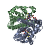
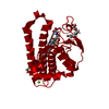
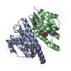
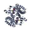
A: Glutathione S-transferase, N-terminal domain protein
B: Glutathione S-transferase, N-terminal domain protein
C: Tripeptide likely a portion of the N-terminal tag
hetero molecules


Theoretical massNumber of molelcules
Total (without water)52,3666
Polymers51,7273
Non-polymers6393
Water7,260403
1
A: Glutathione S-transferase, N-terminal domain protein
hetero molecules

A: Glutathione S-transferase, N-terminal domain protein
hetero molecules


Theoretical massNumber of molelcules
Total (without water)52,0684
Polymers51,4542
Non-polymers6152
Water362
TypeNameSymmetry operationNumber
identity operation1_555x,y,z1
crystal symmetry operation3_656-x+1,y,-z+3/21
Buried area4140 Å2
ΔGint-23 kcal/mol
Surface area16110 Å2
MethodPISA
2
B: Glutathione S-transferase, N-terminal domain protein
hetero molecules

B: Glutathione S-transferase, N-terminal domain protein
hetero molecules


Theoretical massNumber of molelcules
Total (without water)52,1176
Polymers51,4542
Non-polymers6634
Water362
TypeNameSymmetry operationNumber
identity operation1_555x,y,z1
crystal symmetry operation3_555-x,y,-z+1/21
Buried area4260 Å2
ΔGint-16 kcal/mol
Surface area16300 Å2
MethodPISA
3
C: Tripeptide likely a portion of the N-terminal tag


  • defined by author&software
  • 273 Da, 1 polymers
Theoretical massNumber of molelcules
Total (without water)2731
Polymers2731
Non-polymers00
Water181
TypeNameSymmetry operationNumber
identity operation1_555x,y,z1
MethodPISA
Unit cell
Length a, b, c (Å)100.040, 170.730, 50.781
Angle α, β, γ (deg.)90.000, 90.000, 90.000
Int Tables number20
Space group name H-MC2221
Components on special symmetry positions
IDModelComponents
11A-429-

HOH

21A-558-

HOH

31B-621-

HOH

-
Components

#1: Protein Glutathione S-transferase, N-terminal domain protein /


Mass: 25726.910 Da / Num. of mol.: 2
Source method: isolated from a genetically manipulated source
Source: (gene. exp.) Burkholderia ambifaria (bacteria) / Strain: ATCC BAA-244 / AMMD / Gene: Bamb_4793 / Plasmid: pET / Production host: Escherichia coli (E. coli) / Strain (production host): BL21(DE3) / References: UniProt: Q0B681
#2: Protein/peptide Tripeptide likely a portion of the N-terminal tag


Mass: 273.330 Da / Num. of mol.: 1
Source method: isolated from a genetically manipulated source
Source: (gene. exp.) Burkholderia ambifaria (bacteria) / Strain: ATCC BAA-244 / AMMD / Gene: Bamb_4793 / Plasmid: pET / Production host: Escherichia coli (E. coli) / Strain (production host): BL21(DE3)
#3: Chemical ChemComp-GSH / GLUTATHIONE / Glutathione


Mass: 307.323 Da / Num. of mol.: 2 / Source method: obtained synthetically / Formula: C10H17N3O6S
#4: Chemical ChemComp-MG / MAGNESIUM ION


Mass: 24.305 Da / Num. of mol.: 1 / Source method: obtained synthetically / Formula: Mg
#5: Water ChemComp-HOH / water / Water


Mass: 18.015 Da / Num. of mol.: 403 / Source method: isolated from a natural source / Formula: H2O
Compound detailsAUTHORS STATE THAT THE TRIPEPTIDE IS MOST LIKELY A PORTION OF THE N-TERMINAL TAG, BUT IT WAS NOT ...AUTHORS STATE THAT THE TRIPEPTIDE IS MOST LIKELY A PORTION OF THE N-TERMINAL TAG, BUT IT WAS NOT DEFINED ENOUGH TO DEFINITIVELY DEFINE WHICH THREE RESIDUES FROM THE TAG THEY WERE OR EVEN WHICH SUBUNIT IT WAS COMING FROM. COMPLICATING THIS ANALYSIS IS THAT THE TAG IS CLEAVABLE AND TEV WAS INCLUDED IN THE CRYSTALLIZATION SETUP, SO THE TAG MAY BE CLEAVED AND FREE IN SOLUTION.

-
Experimental details

-
Experiment

ExperimentMethod: X-RAY DIFFRACTION / Number of used crystals: 1

-
Sample preparation

CrystalDensity Matthews: 2.1 Å3/Da / Density % sol: 41.31 %
Crystal growTemperature: 298 K / Method: vapor diffusion, sitting drop
Details: Protein (10 mM Hepes pH 7.5, 150 mM NaCl, 5% glycerol). Reservoir (0.2 M MgFormate pH 5.9, 20% peg3350). Cryoprotection (Reservoir + 20% diethylene glycol), VAPOR DIFFUSION, SITTING DROP, temperature 298K

-
Data collection

DiffractionMean temperature: 100 K
Diffraction sourceSource: SYNCHROTRON / Site: APS / Beamline: 31-ID / Wavelength: 0.9793 Å
DetectorType: RAYONIX MX225HE / Detector: CCD / Date: Jul 9, 2012 / Details: MIRRORS
RadiationMonochromator: GRAPHITE / Protocol: SINGLE WAVELENGTH / Monochromatic (M) / Laue (L): M / Scattering type: x-ray
Radiation wavelengthWavelength: 0.9793 Å / Relative weight: 1
ReflectionResolution: 1.6→86.314 Å / Num. all: 57737 / Num. obs: 57737 / % possible obs: 99.9 % / Observed criterion σ(F): 0 / Observed criterion σ(I): 0 / Redundancy: 7.4 % / Rsym value: 0.082 / Net I/σ(I): 11.5
Reflection shell

Diffraction-ID: 1

Resolution (Å)Redundancy (%)Rmerge(I) obsMean I/σ(I) obsNum. measured allNum. unique allRsym value% possible all
1.6-1.697.40.7051.16174283650.705100
1.69-1.797.40.4771.65838778860.477100
1.79-1.917.40.3152.45515674270.315100
1.91-2.077.40.1913.95174669520.191100
2.07-2.267.50.1335.14769863960.133100
2.26-2.537.50.1135.54360458400.113100
2.53-2.927.40.1085.43818351320.108100
2.92-3.587.40.0787.53264343950.078100
3.58-5.067.30.03615.82512334400.03699.8
5.06-27.3556.80.02919.81297819040.02996.7

-
Processing

Software
NameVersionClassificationNB
SCALA3.3.20data scaling
PHENIX1.8.1_1168refinement
PDB_EXTRACT3.11data extraction
MOSFLMdata reduction
BALBESphasing
RefinementMethod to determine structure: MOLECULAR REPLACEMENT
Starting model: 3M3M
Resolution: 1.6→27.355 Å / Occupancy max: 1 / Occupancy min: 0.3 / FOM work R set: 0.8451 / SU ML: 0.16 / σ(F): 0 / σ(I): 0 / Phase error: 22.3 / Stereochemistry target values: ML
RfactorNum. reflection% reflectionSelection details
Rfree0.2162 2921 5.06 %RANDOM
Rwork0.1865 ---
obs0.188 57709 99.84 %-
all-57709 --
Solvent computationShrinkage radii: 0.9 Å / VDW probe radii: 1.11 Å / Solvent model: FLAT BULK SOLVENT MODEL
Displacement parametersBiso max: 78.47 Å2 / Biso mean: 28.7031 Å2 / Biso min: 7.37 Å2
Refinement stepCycle: LAST / Resolution: 1.6→27.355 Å
ProteinNucleic acidLigandSolventTotal
Num. atoms3222 0 41 403 3666
Refine LS restraints
Refine-IDTypeDev idealNumber
X-RAY DIFFRACTIONf_bond_d0.0073378
X-RAY DIFFRACTIONf_angle_d1.0764607
X-RAY DIFFRACTIONf_chiral_restr0.077478
X-RAY DIFFRACTIONf_plane_restr0.005608
X-RAY DIFFRACTIONf_dihedral_angle_d14.2841229
LS refinement shell

Refine-ID: X-RAY DIFFRACTION / Total num. of bins used: 21

Resolution (Å)Rfactor RfreeNum. reflection RfreeRfactor RworkNum. reflection RworkNum. reflection all% reflection obs (%)
1.6-1.62620.291380.245525822720100
1.6262-1.65430.26381350.238325792714100
1.6543-1.68430.25971600.221825682728100
1.6843-1.71670.25451070.222125882695100
1.7167-1.75180.27981400.223326002740100
1.7518-1.78990.23741470.208625562703100
1.7899-1.83150.24171440.207125732717100
1.8315-1.87730.2541390.204326242763100
1.8773-1.9280.22081160.220625912707100
1.928-1.98470.20821360.201226012737100
1.9847-2.04880.22851460.189325812727100
2.0488-2.1220.22511570.190825832740100
2.122-2.20690.25631370.185426012738100
2.2069-2.30730.26741510.193426122763100
2.3073-2.42890.22181450.178625982743100
2.4289-2.58090.211410.181826142755100
2.5809-2.780.22161490.179426152764100
2.78-3.05950.20661350.180726212756100
3.0595-3.50140.20561380.180626622800100
3.5014-4.40840.16511290.156926922821100
4.4084-27.35890.20081310.18892747287897
Refinement TLS params.

Method: refined / Refine-ID: X-RAY DIFFRACTION

IDL112)L122)L132)L222)L232)L332)S11 (Å °)S12 (Å °)S13 (Å °)S21 (Å °)S22 (Å °)S23 (Å °)S31 (Å °)S32 (Å °)S33 (Å °)T112)T122)T132)T222)T232)T332)Origin x (Å)Origin y (Å)Origin z (Å)
10.08820.0069-0.00050.0555-0.06120.0731-0.0957-0.06980.3198-0.0311-0.00010.0133-0.0836-0.0392-0.03550.09810.0407-0.06030.1403-0.07550.171241.842459.959745.529
20.0803-0.0203-0.07210.07190.02760.07670.11910.10670.3972-0.13390.03790.0703-0.2765-0.13610.18960.15850.1093-0.11510.1330.12410.419734.843370.856231.5356
30.1095-0.05460.0640.13480.04250.06740.01830.10590.088-0.0186-0.0391-0.1478-0.0064-0.0045-0.00130.0980.00630.00770.14130.01750.11111.421456.74249.7147
40.2234-0.1216-0.08080.09570.05140.06750.0853-0.07860.37370.21780.0788-0.3688-0.11560.05740.05610.19410.0122-0.11950.1072-0.07940.16569.430467.309724.8467
50.03420.0532-0.01250.2667-0.06590.0523-0.0022-0.00110.0010.004-0.0042-00.0005-0.0026-0.00070.37230.03740.00170.4068-0.01260.460827.213255.25519.2823
Refinement TLS group
IDRefine-IDRefine TLS-IDSelection detailsAuth asym-IDAuth seq-ID
1X-RAY DIFFRACTION1CHAIN A AND (RESID 0:102)A0
2X-RAY DIFFRACTION2CHAIN A AND (RESID 103:207)A0
3X-RAY DIFFRACTION3CHAIN B AND (RESID 2:101)B0
4X-RAY DIFFRACTION4CHAIN B AND (RESID 102:207)B0
5X-RAY DIFFRACTION5CHAIN C AND (RESID 332:334)C0

+
About Yorodumi

-
News

-
Feb 9, 2022. New format data for meta-information of EMDB entries

New format data for meta-information of EMDB entries

  • Version 3 of the EMDB header file is now the official format.
  • The previous official version 1.9 will be removed from the archive.

Related info.:EMDB header

External links:wwPDB to switch to version 3 of the EMDB data model

-
Aug 12, 2020. Covid-19 info

Covid-19 info

URL: https://pdbj.org/emnavi/covid19.php

New page: Covid-19 featured information page in EM Navigator.

Related info.:Covid-19 info / Mar 5, 2020. Novel coronavirus structure data

+
Mar 5, 2020. Novel coronavirus structure data

Novel coronavirus structure data

Related info.:Yorodumi Speices / Aug 12, 2020. Covid-19 info

External links:COVID-19 featured content - PDBj / Molecule of the Month (242):Coronavirus Proteases

+
Jan 31, 2019. EMDB accession codes are about to change! (news from PDBe EMDB page)

EMDB accession codes are about to change! (news from PDBe EMDB page)

  • The allocation of 4 digits for EMDB accession codes will soon come to an end. Whilst these codes will remain in use, new EMDB accession codes will include an additional digit and will expand incrementally as the available range of codes is exhausted. The current 4-digit format prefixed with “EMD-” (i.e. EMD-XXXX) will advance to a 5-digit format (i.e. EMD-XXXXX), and so on. It is currently estimated that the 4-digit codes will be depleted around Spring 2019, at which point the 5-digit format will come into force.
  • The EM Navigator/Yorodumi systems omit the EMD- prefix.

Related info.:Q: What is EMD? / ID/Accession-code notation in Yorodumi/EM Navigator

External links:EMDB Accession Codes are Changing Soon! / Contact to PDBj

+
Jul 12, 2017. Major update of PDB

Major update of PDB

  • wwPDB released updated PDB data conforming to the new PDBx/mmCIF dictionary.
  • This is a major update changing the version number from 4 to 5, and with Remediation, in which all the entries are updated.
  • In this update, many items about electron microscopy experimental information are reorganized (e.g. em_software).
  • Now, EM Navigator and Yorodumi are based on the updated data.

External links:wwPDB Remediation / Enriched Model Files Conforming to OneDep Data Standards Now Available in the PDB FTP Archive

-
Yorodumi

Thousand views of thousand structures

  • Yorodumi is a browser for structure data from EMDB, PDB, SASBDB, etc.
  • This page is also the successor to EM Navigator detail page, and also detail information page/front-end page for Omokage search.
  • The word "yorodu" (or yorozu) is an old Japanese word meaning "ten thousand". "mi" (miru) is to see.

Related info.:EMDB / PDB / SASBDB / Comparison of 3 databanks / Yorodumi Search / Aug 31, 2016. New EM Navigator & Yorodumi / Yorodumi Papers / Jmol/JSmol / Function and homology information / Changes in new EM Navigator and Yorodumi

Read more