 Movie
Movie Controller

[English] 日本語
 Yorodumi
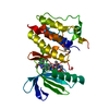
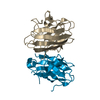
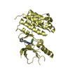
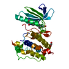
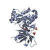
- PDB-4i5i: Crystal structure of the SIRT1 catalytic domain bound to NAD and ... -
+ Open data

- Basic information
| Entry | Database: PDB / ID: 4i5i | ||||||
|---|---|---|---|---|---|---|---|
| Title | Crystal structure of the SIRT1 catalytic domain bound to NAD and an EX527 analog | ||||||
 Components Components | NAD-dependent protein deacetylase sirtuin-1 | ||||||
 Keywords Keywords |  HYDROLASE / Rossmann Fold / histone deacetylase / epigenetics / cancer / sirtuin / acetylated lysine of histone HYDROLASE / Rossmann Fold / histone deacetylase / epigenetics / cancer / sirtuin / acetylated lysine of histone | ||||||
| Function / homology |  Function and homology information Function and homology informationnegative regulation of prostaglandin biosynthetic process / regulation of smooth muscle cell apoptotic process / maintenance of nucleus location / eNoSc complex / histone H4K12 deacetylase activity / histone H3K deacetylase activity / NAD-dependent histone decrotonylase activity / negative regulation of attachment of mitotic spindle microtubules to kinetochore / negative regulation of cellular response to testosterone stimulus / protein depropionylation ...negative regulation of prostaglandin biosynthetic process / regulation of smooth muscle cell apoptotic process / maintenance of nucleus location / eNoSc complex / histone H4K12 deacetylase activity / histone H3K deacetylase activity / NAD-dependent histone decrotonylase activity / negative regulation of attachment of mitotic spindle microtubules to kinetochore / negative regulation of cellular response to testosterone stimulus / protein depropionylation / protein-propionyllysine depropionylase activity / regulation of peroxisome proliferator activated receptor signaling pathway / regulation of transcription by glucose / regulation of protein serine/threonine kinase activity / positive regulation of macrophage apoptotic process / NAD-dependent histone H3K14 deacetylase activity / negative regulation of triglyceride biosynthetic process / regulation of endodeoxyribonuclease activity / triglyceride mobilization / behavioral response to starvation / pyrimidine dimer repair by nucleotide-excision repair / keratin filament binding / HLH domain binding / regulation of lipid storage / leptin-mediated signaling pathway / NAD-dependent histone H3K9 deacetylase activity / positive regulation of smooth muscle cell differentiation / negative regulation of peptidyl-lysine acetylation / NAD-dependent histone H4K16 deacetylase activity / regulation of brown fat cell differentiation / deacetylase activity / response to leptin / bHLH transcription factor binding / positive regulation of endoplasmic reticulum stress-induced intrinsic apoptotic signaling pathway / intracellular triglyceride homeostasis / peptidyl-lysine acetylation / : / negative regulation of androgen receptor signaling pathway / positive regulation of adaptive immune response / regulation of centrosome duplication / rDNA heterochromatin / ovulation from ovarian follicle / single strand break repair / negative regulation of cAMP-dependent protein kinase activity / regulation of bile acid biosynthetic process / NAD-dependent protein lysine deacetylase activity / rDNA heterochromatin formation / protein acetyllysine N-acetyltransferase / NAD-dependent histone deacetylase activity / negative regulation of protein acetylation / negative regulation of phosphorylation / chromatin silencing complex / negative regulation of TOR signaling / protein deacetylation / DNA methylation-dependent heterochromatin formation / UV-damage excision repair / positive regulation of MHC class II biosynthetic process / protein lysine deacetylase activity / negative regulation of helicase activity / positive regulation of cAMP-dependent protein kinase activity / positive regulation of macrophage cytokine production / mitogen-activated protein kinase binding / DNA repair-dependent chromatin remodeling / positive regulation of double-strand break repair / Regulation of FOXO transcriptional activity by acetylation / nuclear inner membrane / muscle organ development / histone deacetylase activity / stress-induced premature senescence / negative regulation of fat cell differentiation / DNA synthesis involved in DNA repair / negative regulation of intrinsic apoptotic signaling pathway in response to DNA damage by p53 class mediator / negative regulation of NF-kappaB transcription factor activity / intracellular glucose homeostasis / negative regulation of cellular senescence / negative regulation of DNA damage response, signal transduction by p53 class mediator / positive regulation of cysteine-type endopeptidase activity involved in apoptotic process / positive regulation of macroautophagy / macrophage differentiation / white fat cell differentiation / negative regulation of cell cycle / regulation of glucose metabolic process / negative regulation of phosphatidylinositol 3-kinase/protein kinase B signal transduction / NAD+ binding / Regulation of HSF1-mediated heat shock response / positive regulation of cholesterol efflux / intrinsic apoptotic signaling pathway in response to DNA damage by p53 class mediator / positive regulation of blood vessel endothelial cell migration / heterochromatin / cellular response to glucose starvation / fatty acid homeostasis / heterochromatin formation / energy homeostasis / negative regulation of oxidative stress-induced intrinsic apoptotic signaling pathway / regulation of cellular response to heat / negative regulation of canonical NF-kappaB signal transduction / positive regulation of insulin receptor signaling pathway / positive regulation of gluconeogenesis / Transferases; Acyltransferases; Transferring groups other than aminoacyl groups / regulation of mitotic cell cycleSimilarity search - Function | ||||||
| Biological species |  Homo sapiens (human) Homo sapiens (human) | ||||||
| Method | X-RAY DIFFRACTION / SYNCHROTRON / MOLECULAR REPLACEMENT / Resolution: 2.5 Å | ||||||
 Authors Authors | Zhao, X. / Allison, D. / Condon, B. / Zhang, F. / Gheyi, T. / Zhang, A. / Ashok, S. / Russell, M. / Macewan, I. / Qian, Y. ...Zhao, X. / Allison, D. / Condon, B. / Zhang, F. / Gheyi, T. / Zhang, A. / Ashok, S. / Russell, M. / Macewan, I. / Qian, Y. / Jamison, J.A. / Luz, J.G. | ||||||
 Citation Citation | Journal: J.Med.Chem. / Year: 2013 Title: The 2.5 angstrom crystal structure of the SIRT1 catalytic domain bound to nicotinamide adenine dinucleotide (NAD+) and an indole (EX527 analogue) reveals a novel mechanism of histone deacetylase inhibition. Authors: Zhao, X. / Allison, D. / Condon, B. / Zhang, F. / Gheyi, T. / Zhang, A. / Ashok, S. / Russell, M. / MacEwan, I. / Qian, Y. / Jamison, J.A. / Luz, J.G. | ||||||
| History |
|
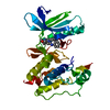
- Structure visualization
| Structure viewer | Molecule: MolmilJmol/JSmol |
|---|
- Downloads & links
-Download
| PDBx/mmCIF format | 4i5i.cif.gz | 125.7 KB | Display | PDBx/mmCIF format |
|---|---|---|---|---|
| PDB format | pdb4i5i.ent.gz | 97.2 KB | Display | PDB format |
| PDBx/mmJSON format | 4i5i.json.gz | Tree view | PDBx/mmJSON format | |
| Others |  Other downloads Other downloads |
-Validation report
| Arichive directory | https://data.pdbj.org/pub/pdb/validation_reports/i5/4i5iftp://data.pdbj.org/pub/pdb/validation_reports/i5/4i5i | HTTPS FTP |
|---|
-Related structure data
| Related structure data | 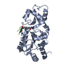 2h2hS S: Starting model for refinement |
|---|---|
| Similar structure data |


-Links
 PDBj
PDBj


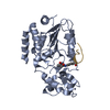
- Assembly
| Deposited unit | 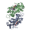
| ||||||||
|---|---|---|---|---|---|---|---|---|---|
| 1 | 
| ||||||||
| 2 | 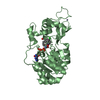
| ||||||||
| 3 |
| ||||||||
| Unit cell |
|
-Components
| #1: Protein | Mass: 32520.723 Da / Num. of mol.: 2 Fragment: Deacetylase sirtuin-type catalytic domain residues 241-516 Source method: isolated from a genetically manipulated source Source: (gene. exp.) Homo sapiens (human) / Gene: SIRT1, SIR2L1 / Cell line (production host): SF9 / Production host:   Spodoptera frugiperda (fall armyworm) Spodoptera frugiperda (fall armyworm)References: UniProt: Q96EB6, Hydrolases; Acting on carbon-nitrogen bonds, other than peptide bonds; In linear amides#2: Chemical | 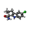  Mass: 262.735 Da / Num. of mol.: 2 / Source method: obtained synthetically / Formula: C14H15ClN2O Mass: 262.735 Da / Num. of mol.: 2 / Source method: obtained synthetically / Formula: C14H15ClN2O#3: Chemical | Nicotinamide adenine dinucleotide  Mass: 663.425 Da / Num. of mol.: 2 / Source method: obtained synthetically / Formula: C21H27N7O14P2 / Comment: NAD*YM Mass: 663.425 Da / Num. of mol.: 2 / Source method: obtained synthetically / Formula: C21H27N7O14P2 / Comment: NAD*YM#4: Chemical |   Mass: 65.409 Da / Num. of mol.: 2 / Source method: obtained synthetically / Formula: Zn Mass: 65.409 Da / Num. of mol.: 2 / Source method: obtained synthetically / Formula: Zn#5: Water | ChemComp-HOH / | Water Mass: 18.015 Da / Num. of mol.: 162 / Source method: isolated from a natural source / Formula: H2O Mass: 18.015 Da / Num. of mol.: 162 / Source method: isolated from a natural source / Formula: H2O |
|---|
-Experimental details
-Experiment
| Experiment | Method: X-RAY DIFFRACTION / Number of used crystals: 1 |
|---|
- Sample preparation
| Crystal | Density Matthews: 4.06 Å3/Da / Density % sol: 69.72 % |
|---|---|
| Crystal grow | Temperature: 294 K / Method: vapor diffusion, hanging drop / pH: 5.4 Details: 300 mM NDSB-195 + 100 mM MES pH 5.4 + 1500 mM Sodium Potassium Tartrate, VAPOR DIFFUSION, HANGING DROP, temperature 294K |
-Data collection
| Diffraction | Mean temperature: 110 K |
|---|---|
| Diffraction source | Source: SYNCHROTRON / Site: APS  / Beamline: 31-ID / Wavelength: 0.97929 Å / Beamline: 31-ID / Wavelength: 0.97929 Å |
| Detector | Type: RAYONIX MX225HE / Detector: CCD / Date: Oct 11, 2009 |
| Radiation | Monochromator: Kohzu HLD-4 Double Crystal / Protocol: SINGLE WAVELENGTH / Monochromatic (M) / Laue (L): M / Scattering type: x-ray |
| Radiation wavelength | Wavelength: 0.97929 Å / Relative weight: 1 |
| Reflection | Resolution: 2.5→32.2 Å / Num. all: 35389 / Num. obs: 35389 / % possible obs: 100 % / Observed criterion σ(F): 1 / Observed criterion σ(I): 1 |
| Reflection shell | Resolution: 2.5→2.64 Å / % possible all: 100 |
- Processing
| Software |
| ||||||||||||||||||||||||||||||||||||||||||||||||||||||||||||||||||||||||||||||||||||||||||||||||||||||||||||||||||||||||||||||||||||||||||||||||||||||||||||||||||||||||||
|---|---|---|---|---|---|---|---|---|---|---|---|---|---|---|---|---|---|---|---|---|---|---|---|---|---|---|---|---|---|---|---|---|---|---|---|---|---|---|---|---|---|---|---|---|---|---|---|---|---|---|---|---|---|---|---|---|---|---|---|---|---|---|---|---|---|---|---|---|---|---|---|---|---|---|---|---|---|---|---|---|---|---|---|---|---|---|---|---|---|---|---|---|---|---|---|---|---|---|---|---|---|---|---|---|---|---|---|---|---|---|---|---|---|---|---|---|---|---|---|---|---|---|---|---|---|---|---|---|---|---|---|---|---|---|---|---|---|---|---|---|---|---|---|---|---|---|---|---|---|---|---|---|---|---|---|---|---|---|---|---|---|---|---|---|---|---|---|---|---|---|---|
| Refinement | Method to determine structure: MOLECULAR REPLACEMENT Starting model: PDB ENTRY 2H2H Resolution: 2.5→30 Å / Cor.coef. Fo:Fc: 0.951 / Cor.coef. Fo:Fc free: 0.925 / SU B: 5.201 / SU ML: 0.117 / Cross valid method: THROUGHOUT / ESU R: 0.23 / ESU R Free: 0.182 / Stereochemistry target values: MAXIMUM LIKELIHOOD
| ||||||||||||||||||||||||||||||||||||||||||||||||||||||||||||||||||||||||||||||||||||||||||||||||||||||||||||||||||||||||||||||||||||||||||||||||||||||||||||||||||||||||||
| Solvent computation | Ion probe radii: 0.8 Å / Shrinkage radii: 0.8 Å / VDW probe radii: 1.2 Å / Solvent model: MASK | ||||||||||||||||||||||||||||||||||||||||||||||||||||||||||||||||||||||||||||||||||||||||||||||||||||||||||||||||||||||||||||||||||||||||||||||||||||||||||||||||||||||||||
| Displacement parameters | Biso mean: 35.601 Å2
| ||||||||||||||||||||||||||||||||||||||||||||||||||||||||||||||||||||||||||||||||||||||||||||||||||||||||||||||||||||||||||||||||||||||||||||||||||||||||||||||||||||||||||
| Refinement step | Cycle: LAST / Resolution: 2.5→30 Å
| ||||||||||||||||||||||||||||||||||||||||||||||||||||||||||||||||||||||||||||||||||||||||||||||||||||||||||||||||||||||||||||||||||||||||||||||||||||||||||||||||||||||||||
| Refine LS restraints |
| ||||||||||||||||||||||||||||||||||||||||||||||||||||||||||||||||||||||||||||||||||||||||||||||||||||||||||||||||||||||||||||||||||||||||||||||||||||||||||||||||||||||||||
| LS refinement shell | Resolution: 2.5→2.565 Å / Total num. of bins used: 20
|
