 Movie
Movie Controller


+ Open data

- Basic information
| Entry | Database: PDB / ID: 4i58 | ||||||
|---|---|---|---|---|---|---|---|
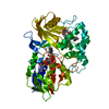
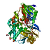
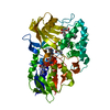
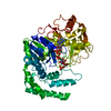
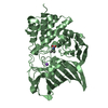
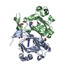
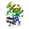
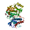
| Title | Cyclohexylamine Oxidase from Brevibacterium oxydans IH-35A | ||||||
 Components Components | Cyclohexylamine Oxidase | ||||||
 Keywords Keywords | OXIDOREDUCTASE / flavoprotein / monoamine oxidase / cyclohexylamine oxidase / biocatalysis | ||||||
| Function / homology |  Function and homology information Function and homology information | ||||||
| Biological species |  Brevibacterium oxydans (bacteria) Brevibacterium oxydans (bacteria) | ||||||
| Method | X-RAY DIFFRACTION / SYNCHROTRON / MOLECULAR REPLACEMENT / Resolution: 3 Å | ||||||
 Authors Authors | Mirza, I.A. / Berghuis, A.M. | ||||||
 Citation Citation | Journal: Plos One / Year: 2013 Title: Structural Analysis of a Novel Cyclohexylamine Oxidase from Brevibacterium oxydans IH-35A. Authors: Mirza, I.A. / Burk, D.L. / Xiong, B. / Iwaki, H. / Hasegawa, Y. / Grosse, S. / Lau, P.C. / Berghuis, A.M. | ||||||
| History |
|
- Structure visualization
| Structure viewer | Molecule: MolmilJmol/JSmol |
|---|
- Downloads & links
-Download
| PDBx/mmCIF format | 4i58.cif.gz | 343.8 KB | Display | PDBx/mmCIF format |
|---|---|---|---|---|
| PDB format | pdb4i58.ent.gz | 282.2 KB | Display | PDB format |
| PDBx/mmJSON format | 4i58.json.gz | Tree view | PDBx/mmJSON format | |
| Others |  Other downloads Other downloads |
-Validation report
| Arichive directory | https://data.pdbj.org/pub/pdb/validation_reports/i5/4i58ftp://data.pdbj.org/pub/pdb/validation_reports/i5/4i58 | HTTPS FTP |
|---|
-Related structure data
| Related structure data | 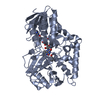 4i59C 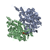 1gosS C: citing same article ( S: Starting model for refinement |
|---|---|
| Similar structure data |


-Links
 PDBj
PDBj
- Assembly
| Deposited unit | 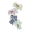
| ||||||||||||||||||||
|---|---|---|---|---|---|---|---|---|---|---|---|---|---|---|---|---|---|---|---|---|---|
| 1 | 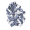
| ||||||||||||||||||||
| 2 | 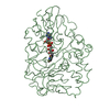
| ||||||||||||||||||||
| 3 | 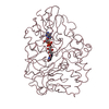
| ||||||||||||||||||||
| 4 | 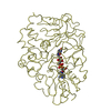
| ||||||||||||||||||||
| Unit cell |
| ||||||||||||||||||||
| Noncrystallographic symmetry (NCS) | NCS domain: (Details: chain AAAA,CCCC, using strict) NCS oper:
|
-Components
| #1: Protein | Mass: 51139.906 Da / Num. of mol.: 4 Source method: isolated from a genetically manipulated source Source: (gene. exp.) Brevibacterium oxydans (bacteria) / Strain: IH-35A / Gene: chaA / Plasmid: pSD80 / Production host: Escherichia coli (E. coli) / Strain (production host): BL21 / References: UniProt: R4GRV2*PLUS#2: Chemical | ChemComp-FAD / Flavin adenine dinucleotide  Mass: 785.550 Da / Num. of mol.: 4 / Source method: obtained synthetically / Formula: C27H33N9O15P2 / Comment: FAD*YM Mass: 785.550 Da / Num. of mol.: 4 / Source method: obtained synthetically / Formula: C27H33N9O15P2 / Comment: FAD*YM |
|---|
-Experimental details
-Experiment
| Experiment | Method: X-RAY DIFFRACTION / Number of used crystals: 1 |
|---|
- Sample preparation
| Crystal | Density Matthews: 2.93 Å3/Da / Density % sol: 57.64 % |
|---|---|
| Crystal grow | Temperature: 298 K / Method: vapor diffusion / pH: 6.5 Details: 30% PEG 8000, 0.2 M sodium acetate trihydrate, 0.1 M sodium cacodylate, pH 6.5, VAPOR DIFFUSION, temperature 298K |
-Data collection
| Diffraction | Mean temperature: 93 K | ||||||||||||||||||||||||||||||||||||||||||||
|---|---|---|---|---|---|---|---|---|---|---|---|---|---|---|---|---|---|---|---|---|---|---|---|---|---|---|---|---|---|---|---|---|---|---|---|---|---|---|---|---|---|---|---|---|---|
| Diffraction source | Source: SYNCHROTRON / Site: NSLS  / Beamline: X8C / Wavelength: 1.1 Å / Beamline: X8C / Wavelength: 1.1 Å | ||||||||||||||||||||||||||||||||||||||||||||
| Detector | Type: ADSC QUANTUM 4r / Detector: CCD / Details: Mirrors | ||||||||||||||||||||||||||||||||||||||||||||
| Radiation | Monochromator: Si (111) / Protocol: SINGLE WAVELENGTH / Monochromatic (M) / Laue (L): M / Scattering type: x-ray | ||||||||||||||||||||||||||||||||||||||||||||
| Radiation wavelength | Wavelength: 1.1 Å / Relative weight: 1 | ||||||||||||||||||||||||||||||||||||||||||||
| Reflection | Resolution: 3→50 Å / Num. all: 46346 / Num. obs: 44399 / % possible obs: 95.8 % / Observed criterion σ(F): 0 / Observed criterion σ(I): 0 / Rmerge(I) obs: 0.143 / Net I/σ(I): 8 | ||||||||||||||||||||||||||||||||||||||||||||
| Reflection shell |
|
- Processing
| Software |
| ||||||||||||||||||||||||||||||||||||||||||||||||||||||||||||
|---|---|---|---|---|---|---|---|---|---|---|---|---|---|---|---|---|---|---|---|---|---|---|---|---|---|---|---|---|---|---|---|---|---|---|---|---|---|---|---|---|---|---|---|---|---|---|---|---|---|---|---|---|---|---|---|---|---|---|---|---|---|
| Refinement | Method to determine structure: MOLECULAR REPLACEMENT Starting model: PDB ENTRY 1GOS Resolution: 3→45.8 Å / Rfactor Rfree error: 0.004 / Occupancy max: 1 / Occupancy min: 1 / Data cutoff high absF: 85358.23 / Data cutoff low absF: 0 / Isotropic thermal model: RESTRAINED / Cross valid method: THROUGHOUT / σ(F): 0 / Stereochemistry target values: Engh & Huber / Details: BULK SOLVENT MODEL USED
| ||||||||||||||||||||||||||||||||||||||||||||||||||||||||||||
| Solvent computation | Solvent model: FLAT MODEL / Bsol: 24.785 Å2 / ksol: 0.35 e/Å3 | ||||||||||||||||||||||||||||||||||||||||||||||||||||||||||||
| Displacement parameters | Biso mean: 26.4 Å2
| ||||||||||||||||||||||||||||||||||||||||||||||||||||||||||||
| Refine analyze |
| ||||||||||||||||||||||||||||||||||||||||||||||||||||||||||||
| Refinement step | Cycle: LAST / Resolution: 3→45.8 Å
| ||||||||||||||||||||||||||||||||||||||||||||||||||||||||||||
| Refine LS restraints |
| ||||||||||||||||||||||||||||||||||||||||||||||||||||||||||||
| Refine LS restraints NCS | NCS model details: CONSTR | ||||||||||||||||||||||||||||||||||||||||||||||||||||||||||||
| LS refinement shell | Resolution: 3→3.19 Å / Rfactor Rfree error: 0.015 / Total num. of bins used: 6
| ||||||||||||||||||||||||||||||||||||||||||||||||||||||||||||
| Xplor file |
|
