 Movie
Movie Controller

[English] 日本語
 Yorodumi
- PDB-4i38: Structures of IT intermediates from time-resolved laue crystallog... -
+ Open data

- Basic information
| Entry | Database: PDB / ID: 4i38 | ||||||
|---|---|---|---|---|---|---|---|
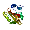
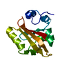
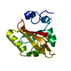
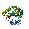
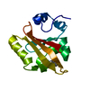
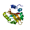
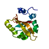
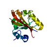
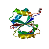
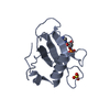
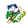
| Title | Structures of IT intermediates from time-resolved laue crystallography collected at 14ID-B, APS | ||||||
 Components Components | Photoactive yellow protein | ||||||
 Keywords Keywords | LUMINESCENT PROTEIN / PHOTORECEPTOR / CHROMOPHORE / PHOTORECEPTOR PROTEIN / RECEPTOR / SENSORY TRANSDUCTION | ||||||
| Function / homology |  Function and homology informationphotoreceptor activity / phototransduction / regulation of DNA-templated transcription / identical protein binding Function and homology informationphotoreceptor activity / phototransduction / regulation of DNA-templated transcription / identical protein bindingSimilarity search - Function | ||||||
| Biological species |  HALORHODOSPIRA HALOPHILA (bacteria) HALORHODOSPIRA HALOPHILA (bacteria) | ||||||
| Method | X-RAY DIFFRACTION / SYNCHROTRON / MOLECULAR REPLACEMENT / Resolution: 1.6 Å | ||||||
 Authors Authors | Jung, Y.O. / Lee, J.H. / Kim, J. / Schmidt, M. / Vukica, S. / Moffat, K. / Ihee, H. | ||||||
 Citation Citation | Journal: NAT.CHEM. / Year: 2013 Title: Volume-conserving trans-cis isomerization pathways in photoactive yellow protein visualized by picosecond X-ray crystallography Authors: Jung, Y.O. / Lee, J.H. / Kim, J. / Schmidt, M. / Moffat, K. / Srajer, V. / Ihee, H. | ||||||
| History |
|
- Structure visualization
| Structure viewer | Molecule: MolmilJmol/JSmol |
|---|
- Downloads & links
-Download
| PDBx/mmCIF format | 4i38.cif.gz | 37.7 KB | Display | PDBx/mmCIF format |
|---|---|---|---|---|
| PDB format | pdb4i38.ent.gz | 24.5 KB | Display | PDB format |
| PDBx/mmJSON format | 4i38.json.gz | Tree view | PDBx/mmJSON format | |
| Others |  Other downloads Other downloads |
-Validation report
| Arichive directory | https://data.pdbj.org/pub/pdb/validation_reports/i3/4i38ftp://data.pdbj.org/pub/pdb/validation_reports/i3/4i38 | HTTPS FTP |
|---|
-Related structure data
| Related structure data | 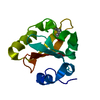 3ve3C 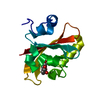 3ve4C 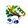 4hy8C 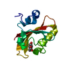 4i39C 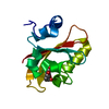 4i3aC  4i3iC 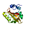 4i3jC 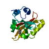 2phyS C: citing same article ( S: Starting model for refinement |
|---|---|
| Similar structure data |


-Links
 PDBj
PDBj


- Assembly
| Deposited unit | 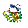
| ||||||||
|---|---|---|---|---|---|---|---|---|---|
| 1 |
| ||||||||
| Unit cell |
|
-Components
| #1: Protein | / PYP Mass: 13888.575 Da / Num. of mol.: 1 Source method: isolated from a genetically manipulated source Source: (gene. exp.) HALORHODOSPIRA HALOPHILA (bacteria) / Gene: pyp / Plasmid: PQE80 / Production host: ESCHERICHIA COLI (E. coli) / Strain (production host): M15 / References: UniProt: P16113 |
|---|---|
| #2: Chemical | ChemComp-HC4 / P-Coumaric acid  Mass: 164.158 Da / Num. of mol.: 1 / Source method: obtained synthetically / Formula: C9H8O3 Mass: 164.158 Da / Num. of mol.: 1 / Source method: obtained synthetically / Formula: C9H8O3 |
-Experimental details
-Experiment
| Experiment | Method: X-RAY DIFFRACTION / Number of used crystals: 1 |
|---|
- Sample preparation
| Crystal | Density Matthews: 1.9 Å3/Da / Density % sol: 35.29 % |
|---|---|
| Crystal grow | Temperature: 298 K / Method: hanging drop / pH: 7 Details: 2.6M AMMONIUM SULFATE, 50MM SODIUM PHOSPHATE, pH 7.0, hanging drop, temperature 298K |
-Data collection
| Diffraction | Mean temperature: 288 K | |||||||||
|---|---|---|---|---|---|---|---|---|---|---|
| Diffraction source | Source: SYNCHROTRON / Site: APS  / Beamline: 14-ID-B / Wavelength: 0.96-1.30 / Beamline: 14-ID-B / Wavelength: 0.96-1.30 | |||||||||
| Detector | Type: MAR CCD 165 mm / Detector: CCD / Date: Nov 17, 2008 | |||||||||
| Radiation | Protocol: LAUE / Monochromatic (M) / Laue (L): L / Scattering type: x-ray | |||||||||
| Radiation wavelength |
| |||||||||
| Reflection | Resolution: 1.6→28.9 Å / Num. obs: 14623 |
- Processing
| Software |
| |||||||||||||||||||||||||||||||||
|---|---|---|---|---|---|---|---|---|---|---|---|---|---|---|---|---|---|---|---|---|---|---|---|---|---|---|---|---|---|---|---|---|---|---|
| Refinement | Method to determine structure: MOLECULAR REPLACEMENT Starting model: PDB ENTRY 2PHY Resolution: 1.6→10 Å / Num. parameters: 3952 / Num. restraintsaints: 4028 / Occupancy max: 1 / Occupancy min: 1 / Cross valid method: FREE R / σ(F): 2 / Stereochemistry target values: ENGH AND HUBER Details: THE STRUCTURE FACTOR FILE REGARDING TO PDB FILE WAS BACK FOURIER-TRANSFORMED AND WAS EXTRAPOLATED FROM THE TIME-INDEPENDENT DIFFERENCE ELECTRON DENSITY MAP. THIS MAP WAS GENERATED USING ...Details: THE STRUCTURE FACTOR FILE REGARDING TO PDB FILE WAS BACK FOURIER-TRANSFORMED AND WAS EXTRAPOLATED FROM THE TIME-INDEPENDENT DIFFERENCE ELECTRON DENSITY MAP. THIS MAP WAS GENERATED USING KINETIC ANALYSIS BASED ON SEVERAL EXPERIMENTAL TIME-DEPENDENT DIFFERENCE ELECTRON DENSITY MAPS.
| |||||||||||||||||||||||||||||||||
| Solvent computation | Solvent model: MOEWS & KRETSINGER, J.MOL.BIOL.91(1973)201-228 | |||||||||||||||||||||||||||||||||
| Displacement parameters | Biso max: 131.06 Å2 / Biso mean: 18.2623 Å2 / Biso min: 2.04 Å2 | |||||||||||||||||||||||||||||||||
| Refinement step | Cycle: LAST / Resolution: 1.6→10 Å
| |||||||||||||||||||||||||||||||||
| Refine LS restraints |
|
