 Movie
Movie Controller

[English] 日本語
 Yorodumi
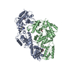
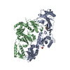
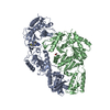
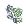
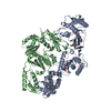
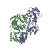
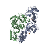
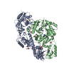
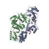
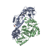
- PDB-4i2p: Crystal structure of HIV-1 reverse transcriptase in complex with ... -
+ Open data

- Basic information
| Entry | Database: PDB / ID: 4i2p | ||||||
|---|---|---|---|---|---|---|---|
| Title | Crystal structure of HIV-1 reverse transcriptase in complex with rilpivirine (TMC278) based analogue | ||||||
 Components Components | (Gag-Pol polyprotein) x 2 | ||||||
 Keywords Keywords |  HYDROLASE / TRANSFERASE/INHIBITOR / P51/P66 / hetero dimer / NNRTI / nonnucleoside inhibitor / AIDS / HIV / DNA recombination / RNA-directed DNA polymerase / DNA polymerase / endonuclease / multifunctional enzyme / transferase / TRANSFERASE-INHIBITOR complex HYDROLASE / TRANSFERASE/INHIBITOR / P51/P66 / hetero dimer / NNRTI / nonnucleoside inhibitor / AIDS / HIV / DNA recombination / RNA-directed DNA polymerase / DNA polymerase / endonuclease / multifunctional enzyme / transferase / TRANSFERASE-INHIBITOR complex | ||||||
| Function / homology |  Function and homology information Function and homology information: / : / HIV-1 retropepsin / retroviral ribonuclease H / exoribonuclease H / exoribonuclease H activity / host multivesicular body / DNA integration / RNA-directed DNA polymerase / viral genome integration into host DNA ...: / : / HIV-1 retropepsin / retroviral ribonuclease H / exoribonuclease H / exoribonuclease H activity / host multivesicular body / DNA integration / RNA-directed DNA polymerase / viral genome integration into host DNA / viral penetration into host nucleus / establishment of integrated proviral latency / RNA-directed DNA polymerase activity / RNA-DNA hybrid ribonuclease activity / Transferases; Transferring phosphorus-containing groups; Nucleotidyltransferases / symbiont-mediated suppression of host gene expression / viral nucleocapsid / DNA recombination / Hydrolases; Acting on ester bonds / DNA-directed DNA polymerase / aspartic-type endopeptidase activity / DNA-directed DNA polymerase activity / symbiont entry into host cell / lipid binding / host cell nucleus / host cell plasma membrane / virion membrane / structural molecule activity / proteolysis / DNA binding / RNA binding / zinc ion binding / membraneSimilarity search - Function | ||||||
| Biological species |  Human immunodeficiency virus type 1 BH10 Human immunodeficiency virus type 1 BH10 | ||||||
| Method | X-RAY DIFFRACTION / SYNCHROTRON / MOLECULAR REPLACEMENT / Resolution: 2.2964 Å | ||||||
 Authors Authors | Patel, D. / Bauman, J.D. / Das, K. / Arnold, E. | ||||||
 Citation Citation | Journal: Retrovirology / Year: 2012 Title: A comparison of the ability of rilpivirine (TMC278) and selected analogues to inhibit clinically relevant HIV-1 reverse transcriptase mutants. Authors: Johnson, B.C. / Pauly, G.T. / Rai, G. / Patel, D. / Bauman, J.D. / Baker, H.L. / Das, K. / Schneider, J.P. / Maloney, D.J. / Arnold, E. / Thomas, C.J. / Hughes, S.H. | ||||||
| History |
|
- Structure visualization
| Structure viewer | Molecule: MolmilJmol/JSmol |
|---|
- Downloads & links
-Download
| PDBx/mmCIF format | 4i2p.cif.gz | 215.9 KB | Display | PDBx/mmCIF format |
|---|---|---|---|---|
| PDB format | pdb4i2p.ent.gz | 170.5 KB | Display | PDB format |
| PDBx/mmJSON format | 4i2p.json.gz | Tree view | PDBx/mmJSON format | |
| Others |  Other downloads Other downloads |
-Validation report
| Arichive directory | https://data.pdbj.org/pub/pdb/validation_reports/i2/4i2pftp://data.pdbj.org/pub/pdb/validation_reports/i2/4i2p | HTTPS FTP |
|---|
-Related structure data


-Links
 PDBj
PDBj


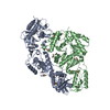
- Assembly
| Deposited unit | 
| ||||||||
|---|---|---|---|---|---|---|---|---|---|
| 1 |
| ||||||||
| Unit cell |
|
-Components
| #1: Protein | Mass: 63989.238 Da / Num. of mol.: 1 / Fragment: p66 (UNP Residues 600-1154) / Mutation: C879S, K771A, K772A Source method: isolated from a genetically manipulated source Source: (gene. exp.) Human immunodeficiency virus type 1 BH10Gene: gag-pol / Production host:  Escherichia coli (E. coli) Escherichia coli (E. coli)References: UniProt: P03366, HIV-1 retropepsin, RNA-directed DNA polymerase, DNA-directed DNA polymerase, retroviral ribonuclease H, exoribonuclease H |
|---|---|
| #2: Protein | Mass: 50039.488 Da / Num. of mol.: 1 / Fragment: p51 (UNP Residues 600-1077) / Mutation: C879S Source method: isolated from a genetically manipulated source Source: (gene. exp.) Human immunodeficiency virus type 1 BH10Gene: gag-pol / Production host: Escherichia coli (E. coli)References: UniProt: P03366, HIV-1 retropepsin, RNA-directed DNA polymerase, DNA-directed DNA polymerase, retroviral ribonuclease H, exoribonuclease H |
| #3: Chemical | ChemComp-G73 / (  Mass: 411.459 Da / Num. of mol.: 1 / Source method: obtained synthetically / Formula: C23H21N7O Mass: 411.459 Da / Num. of mol.: 1 / Source method: obtained synthetically / Formula: C23H21N7O |
| #4: Water | ChemComp-HOH / Water Mass: 18.015 Da / Num. of mol.: 300 / Source method: isolated from a natural source / Formula: H2O Mass: 18.015 Da / Num. of mol.: 300 / Source method: isolated from a natural source / Formula: H2O |
-Experimental details
-Experiment
| Experiment | Method: X-RAY DIFFRACTION / Number of used crystals: 1 |
|---|
- Sample preparation
| Crystal | Density Matthews: 2.8 Å3/Da / Density % sol: 56.13 % |
|---|---|
| Crystal grow | Temperature: 301 K / Method: vapor diffusion, hanging drop / pH: 6.6 Details: 50 mM imidazole pH 6.6, 100 mM ammonium sulfate, 15 mM manganese sulfate, 10 mM spermine, 5 mM TCEP, 11% (w/w) PEG 8000, and 5% PEG 400, VAPOR DIFFUSION, HANGING DROP, temperature 301K |
-Data collection
| Diffraction source | Source: SYNCHROTRON / Site: CHESS  / Beamline: F1 / Beamline: F1 |
|---|---|
| Detector | Type: ADSC QUANTUM 270 / Detector: CCD / Date: Jul 13, 2010 |
| Radiation | Protocol: SINGLE WAVELENGTH / Monochromatic (M) / Laue (L): M / Scattering type: x-ray |
| Radiation wavelength | Relative weight: 1 |
| Reflection | Resolution: 2.296→107.73 Å / Num. obs: 55926 |
- Processing
| Software |
| |||||||||||||||||||||||||||||||||||||||||||||||||||||||||||||||||||||||||||||||||||||||||||||||||||||||||
|---|---|---|---|---|---|---|---|---|---|---|---|---|---|---|---|---|---|---|---|---|---|---|---|---|---|---|---|---|---|---|---|---|---|---|---|---|---|---|---|---|---|---|---|---|---|---|---|---|---|---|---|---|---|---|---|---|---|---|---|---|---|---|---|---|---|---|---|---|---|---|---|---|---|---|---|---|---|---|---|---|---|---|---|---|---|---|---|---|---|---|---|---|---|---|---|---|---|---|---|---|---|---|---|---|---|---|
| Refinement | Method to determine structure: MOLECULAR REPLACEMENT / Resolution: 2.2964→41.294 Å / SU ML: 0.21 / σ(F): 1.34 / Phase error: 26.28 / Stereochemistry target values: ML
| |||||||||||||||||||||||||||||||||||||||||||||||||||||||||||||||||||||||||||||||||||||||||||||||||||||||||
| Solvent computation | Shrinkage radii: 0.6 Å / VDW probe radii: 0.9 Å / Solvent model: FLAT BULK SOLVENT MODEL / Bsol: 42.058 Å2 / ksol: 0.39 e/Å3 | |||||||||||||||||||||||||||||||||||||||||||||||||||||||||||||||||||||||||||||||||||||||||||||||||||||||||
| Displacement parameters |
| |||||||||||||||||||||||||||||||||||||||||||||||||||||||||||||||||||||||||||||||||||||||||||||||||||||||||
| Refinement step | Cycle: LAST / Resolution: 2.2964→41.294 Å
| |||||||||||||||||||||||||||||||||||||||||||||||||||||||||||||||||||||||||||||||||||||||||||||||||||||||||
| Refine LS restraints |
| |||||||||||||||||||||||||||||||||||||||||||||||||||||||||||||||||||||||||||||||||||||||||||||||||||||||||
| LS refinement shell |
|
