 Movie
Movie Controller


+ Open data

- Basic information
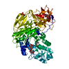
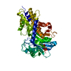
| Entry | Database: PDB / ID: 4i0w | ||||||
|---|---|---|---|---|---|---|---|
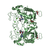
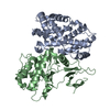
| Title | Structure of the Clostridium Perfringens CspB protease | ||||||
 Components Components | (Protease CspB) x 2 | ||||||
 Keywords Keywords |  HYDROLASE / jellyroll / subtilisin HYDROLASE / jellyroll / subtilisin | ||||||
| Function / homology |  Function and homology informationHydrolases; Acting on peptide bonds (peptidases) / serine-type endopeptidase activity Function and homology informationHydrolases; Acting on peptide bonds (peptidases) / serine-type endopeptidase activitySimilarity search - Function | ||||||
| Biological species |   Clostridium perfringens (bacteria) Clostridium perfringens (bacteria) | ||||||
| Method | X-RAY DIFFRACTION / SYNCHROTRON / SAD / Resolution: 1.6 Å | ||||||
 Authors Authors | Adams, C.M. / Eckenroth, B.E. / Doublie, S. | ||||||
 Citation Citation | Journal: Plos Pathog. / Year: 2013 Title: Structural and functional analysis of the CspB protease required for Clostridium spore germination. Authors: Adams, C.M. / Eckenroth, B.E. / Putnam, E.E. / Doublie, S. / Shen, A. | ||||||
| History |
|
- Structure visualization
| Structure viewer | Molecule: MolmilJmol/JSmol |
|---|
- Downloads & links
-Download
| PDBx/mmCIF format | 4i0w.cif.gz | 460.4 KB | Display | PDBx/mmCIF format |
|---|---|---|---|---|
| PDB format | pdb4i0w.ent.gz | 391.4 KB | Display | PDB format |
| PDBx/mmJSON format | 4i0w.json.gz | Tree view | PDBx/mmJSON format | |
| Others |  Other downloads Other downloads |
-Validation report
| Arichive directory | https://data.pdbj.org/pub/pdb/validation_reports/i0/4i0wftp://data.pdbj.org/pub/pdb/validation_reports/i0/4i0w | HTTPS FTP |
|---|
-Related structure data
| Similar structure data |
|---|


-Links
 PDBj
PDBj

- Assembly
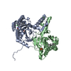
| Deposited unit | 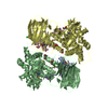
| ||||||||
|---|---|---|---|---|---|---|---|---|---|
| 1 | 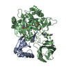
| ||||||||
| 2 | 
| ||||||||
| Unit cell |
|
-Components
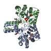
-Protein , 2 types, 4 molecules ACBD
| #1: Protein | Mass: 10855.533 Da / Num. of mol.: 2 / Fragment: prodomain (UNP residues 1-96) Source method: isolated from a genetically manipulated source Source: (gene. exp.) Clostridium perfringens (bacteria) / Gene: CPF_2887, cspB / Plasmid: pRSFduet1-cspB / Production host: Escherichia coli (E. coli) / Strain (production host): BL21(DE3)References: UniProt: Q0TM89, UniProt: A0A0H2YU83*PLUS, Hydrolases; Acting on peptide bonds (peptidases)#2: Protein | Mass: 52855.551 Da / Num. of mol.: 2 / Fragment: subtilase domain (UNP residues 97-565) Source method: isolated from a genetically manipulated source Source: (gene. exp.) Clostridium perfringens (bacteria) / Gene: CPF_2887, cspB / Plasmid: pRSFduet1-cspB / Production host: Escherichia coli (E. coli) / Strain (production host): BL21(DE3)References: UniProt: Q0TM89, UniProt: A0A0H2YU83*PLUS, Hydrolases; Acting on peptide bonds (peptidases) |
|---|
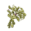
-Non-polymers , 4 types, 959 molecules 


| #3: Chemical | Chloride Mass: 35.453 Da / Num. of mol.: 3 / Source method: obtained synthetically / Formula: Cl Mass: 35.453 Da / Num. of mol.: 3 / Source method: obtained synthetically / Formula: Cl#4: Chemical |  Mass: 22.990 Da / Num. of mol.: 2 / Source method: obtained synthetically / Formula: Na Mass: 22.990 Da / Num. of mol.: 2 / Source method: obtained synthetically / Formula: Na#5: Chemical | ChemComp-EDO / Ethylene glycol Mass: 62.068 Da / Num. of mol.: 8 / Source method: obtained synthetically / Formula: C2H6O2 Mass: 62.068 Da / Num. of mol.: 8 / Source method: obtained synthetically / Formula: C2H6O2#6: Water | ChemComp-HOH / | WaterMass: 18.015 Da / Num. of mol.: 946 / Source method: isolated from a natural source / Formula: H2O |
|---|
-Experimental details
-Experiment
| Experiment | Method: X-RAY DIFFRACTION / Number of used crystals: 1 |
|---|
- Sample preparation
| Crystal | Density Matthews: 2.8 Å3/Da / Density % sol: 56.12 % |
|---|---|
| Crystal grow | Temperature: 285.15 K / Method: vapor diffusion, hanging drop / pH: 5 Details: 27% ethylene glycol, 50 mM sodium acetate, pH 5.0, VAPOR DIFFUSION, HANGING DROP, temperature 285.15K |
-Data collection
| Diffraction | Mean temperature: 100 K |
|---|---|
| Diffraction source | Source: SYNCHROTRON / Site: APS  / Beamline: 23-ID-B / Wavelength: 0.9794 Å / Beamline: 23-ID-B / Wavelength: 0.9794 Å |
| Detector | Type: MARMOSAIC 300 mm CCD / Detector: CCD / Date: Feb 15, 2012 |
| Radiation | Monochromator: double crystal cryo-cooled Si(111) / Protocol: SINGLE WAVELENGTH / Monochromatic (M) / Laue (L): M / Scattering type: x-ray |
| Radiation wavelength | Wavelength: 0.9794 Å / Relative weight: 1 |
| Reflection | Resolution: 1.49→43.735 Å / Num. all: 202733 / Num. obs: 202733 / % possible obs: 86.8 % / Observed criterion σ(I): -3 / Redundancy: 6.6 % / Biso Wilson estimate: 19.1 Å2 / Rmerge(I) obs: 0.078 / Net I/σ(I): 18.419 |
| Reflection shell | Resolution: 1.49→1.54 Å / Redundancy: 2.8 % / Rmerge(I) obs: 0.451 / Mean I/σ(I) obs: 2.05 / % possible all: 46.2 |
-Phasing
| Phasing | Method: SAD |
|---|
- Processing
| Software |
| |||||||||||||||||||||||||||||||||||||||||||||||||||||||||||||||||||||||||||||||||||||||||||||||||||||||||||||||||||||||||||||||||||||||||||||||||||||||||||||||||||||||||||||||||||||||||||||||||||||||||||||||||||||||||
|---|---|---|---|---|---|---|---|---|---|---|---|---|---|---|---|---|---|---|---|---|---|---|---|---|---|---|---|---|---|---|---|---|---|---|---|---|---|---|---|---|---|---|---|---|---|---|---|---|---|---|---|---|---|---|---|---|---|---|---|---|---|---|---|---|---|---|---|---|---|---|---|---|---|---|---|---|---|---|---|---|---|---|---|---|---|---|---|---|---|---|---|---|---|---|---|---|---|---|---|---|---|---|---|---|---|---|---|---|---|---|---|---|---|---|---|---|---|---|---|---|---|---|---|---|---|---|---|---|---|---|---|---|---|---|---|---|---|---|---|---|---|---|---|---|---|---|---|---|---|---|---|---|---|---|---|---|---|---|---|---|---|---|---|---|---|---|---|---|---|---|---|---|---|---|---|---|---|---|---|---|---|---|---|---|---|---|---|---|---|---|---|---|---|---|---|---|---|---|---|---|---|---|---|---|---|---|---|---|---|---|---|---|---|---|---|---|---|---|
| Refinement | Method to determine structure: SAD / Resolution: 1.6→43.735 Å / Occupancy max: 1 / Occupancy min: 0.16 / SU ML: 0.13 / σ(F): 1.34 / Phase error: 18.35 / Stereochemistry target values: MLHL
| |||||||||||||||||||||||||||||||||||||||||||||||||||||||||||||||||||||||||||||||||||||||||||||||||||||||||||||||||||||||||||||||||||||||||||||||||||||||||||||||||||||||||||||||||||||||||||||||||||||||||||||||||||||||||
| Solvent computation | Shrinkage radii: 0.9 Å / VDW probe radii: 1.11 Å / Solvent model: FLAT BULK SOLVENT MODEL | |||||||||||||||||||||||||||||||||||||||||||||||||||||||||||||||||||||||||||||||||||||||||||||||||||||||||||||||||||||||||||||||||||||||||||||||||||||||||||||||||||||||||||||||||||||||||||||||||||||||||||||||||||||||||
| Displacement parameters | Biso mean: 28.4632 Å2 | |||||||||||||||||||||||||||||||||||||||||||||||||||||||||||||||||||||||||||||||||||||||||||||||||||||||||||||||||||||||||||||||||||||||||||||||||||||||||||||||||||||||||||||||||||||||||||||||||||||||||||||||||||||||||
| Refinement step | Cycle: LAST / Resolution: 1.6→43.735 Å
| |||||||||||||||||||||||||||||||||||||||||||||||||||||||||||||||||||||||||||||||||||||||||||||||||||||||||||||||||||||||||||||||||||||||||||||||||||||||||||||||||||||||||||||||||||||||||||||||||||||||||||||||||||||||||
| Refine LS restraints |
| |||||||||||||||||||||||||||||||||||||||||||||||||||||||||||||||||||||||||||||||||||||||||||||||||||||||||||||||||||||||||||||||||||||||||||||||||||||||||||||||||||||||||||||||||||||||||||||||||||||||||||||||||||||||||
| LS refinement shell |
|
