 Movie
Movie Controller

[English] 日本語
 Yorodumi
- PDB-4hzt: Structure-based design of novel dihydroisoquinoline BACE-1 inhibi... -
+ Open data

- Basic information
| Entry | Database: PDB / ID: 4hzt | ||||||
|---|---|---|---|---|---|---|---|
| Title | Structure-based design of novel dihydroisoquinoline BACE-1 inhibitors that do not engage the catalytic aspartates | ||||||
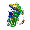
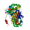
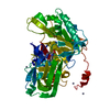
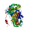
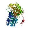
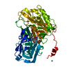
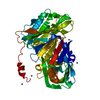
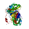
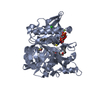
 Components Components | Beta-secretase 1 | ||||||
 Keywords Keywords | hydrolase/hydrolase inhibitor / Aspartic protease / Hydrolysis / hydrolase-hydrolase inhibitor complex | ||||||
| Function / homology |  Function and homology informationmemapsin 2 / Golgi-associated vesicle lumen / signaling receptor ligand precursor processing / beta-aspartyl-peptidase activity / amyloid precursor protein catabolic process / amyloid-beta formation / membrane protein ectodomain proteolysis / cellular response to manganese ion / prepulse inhibition / detection of mechanical stimulus involved in sensory perception of pain ...memapsin 2 / Golgi-associated vesicle lumen / signaling receptor ligand precursor processing / beta-aspartyl-peptidase activity / amyloid precursor protein catabolic process / amyloid-beta formation / membrane protein ectodomain proteolysis / cellular response to manganese ion / prepulse inhibition / detection of mechanical stimulus involved in sensory perception of pain / amyloid-beta metabolic process / cellular response to copper ion / hippocampal mossy fiber to CA3 synapse / presynaptic modulation of chemical synaptic transmission / multivesicular body / response to lead ion / trans-Golgi network / protein processing / recycling endosome / cellular response to amyloid-beta / positive regulation of neuron apoptotic process / synaptic vesicle / late endosome / amyloid-beta binding / peptidase activity / endopeptidase activity / amyloid fibril formation / lysosome / aspartic-type endopeptidase activity / early endosome / endosome membrane / endosome / membrane raft / Amyloid fiber formation / axon / endoplasmic reticulum lumen / neuronal cell body / dendrite / Golgi apparatus / enzyme binding / cell surface / proteolysis / membrane / plasma membrane Function and homology informationmemapsin 2 / Golgi-associated vesicle lumen / signaling receptor ligand precursor processing / beta-aspartyl-peptidase activity / amyloid precursor protein catabolic process / amyloid-beta formation / membrane protein ectodomain proteolysis / cellular response to manganese ion / prepulse inhibition / detection of mechanical stimulus involved in sensory perception of pain ...memapsin 2 / Golgi-associated vesicle lumen / signaling receptor ligand precursor processing / beta-aspartyl-peptidase activity / amyloid precursor protein catabolic process / amyloid-beta formation / membrane protein ectodomain proteolysis / cellular response to manganese ion / prepulse inhibition / detection of mechanical stimulus involved in sensory perception of pain / amyloid-beta metabolic process / cellular response to copper ion / hippocampal mossy fiber to CA3 synapse / presynaptic modulation of chemical synaptic transmission / multivesicular body / response to lead ion / trans-Golgi network / protein processing / recycling endosome / cellular response to amyloid-beta / positive regulation of neuron apoptotic process / synaptic vesicle / late endosome / amyloid-beta binding / peptidase activity / endopeptidase activity / amyloid fibril formation / lysosome / aspartic-type endopeptidase activity / early endosome / endosome membrane / endosome / membrane raft / Amyloid fiber formation / axon / endoplasmic reticulum lumen / neuronal cell body / dendrite / Golgi apparatus / enzyme binding / cell surface / proteolysis / membrane / plasma membraneSimilarity search - Function | ||||||
| Biological species |  Homo sapiens (human) Homo sapiens (human) | ||||||
| Method | X-RAY DIFFRACTION / MOLECULAR REPLACEMENT / Resolution: 1.8 Å | ||||||
 Authors Authors | Yao, N. / Brecht, E. | ||||||
 Citation Citation | Journal: Bioorg.Med.Chem.Lett. / Year: 2013 Title: Structure-based design of novel dihydroisoquinoline BACE-1 inhibitors that do not engage the catalytic aspartates. Authors: Bowers, S. / Xu, Y.Z. / Yuan, S. / Probst, G.D. / Hom, R.K. / Chan, W. / Konradi, A.W. / Sham, H.L. / Zhu, Y.L. / Beroza, P. / Pan, H. / Brecht, E. / Yao, N. / Lougheed, J. / Tam, D. / Ren, ...Authors: Bowers, S. / Xu, Y.Z. / Yuan, S. / Probst, G.D. / Hom, R.K. / Chan, W. / Konradi, A.W. / Sham, H.L. / Zhu, Y.L. / Beroza, P. / Pan, H. / Brecht, E. / Yao, N. / Lougheed, J. / Tam, D. / Ren, Z. / Ruslim, L. / Bova, M.P. / Artis, D.R. | ||||||
| History |
|
- Structure visualization
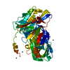
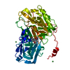
| Structure viewer | Molecule: MolmilJmol/JSmol |
|---|
- Downloads & links
-Download
| PDBx/mmCIF format | 4hzt.cif.gz | 99.3 KB | Display | PDBx/mmCIF format |
|---|---|---|---|---|
| PDB format | pdb4hzt.ent.gz | 79.4 KB | Display | PDB format |
| PDBx/mmJSON format | 4hzt.json.gz | Tree view | PDBx/mmJSON format | |
| Others |  Other downloads Other downloads |
-Validation report
| Arichive directory | https://data.pdbj.org/pub/pdb/validation_reports/hz/4hztftp://data.pdbj.org/pub/pdb/validation_reports/hz/4hzt | HTTPS FTP |
|---|
-Related structure data


-Links
 PDBj
PDBj

- Assembly
| Deposited unit | 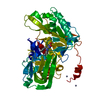
| |||||||||
|---|---|---|---|---|---|---|---|---|---|---|
| 1 |
| |||||||||
| 2 | 
| |||||||||
| Unit cell |
| |||||||||
| Components on special symmetry positions |
|
-Components
| #1: Protein | / Aspartyl protease 2 / ASP2 / Asp 2 / Beta-site amyloid precursor protein cleaving enzyme 1 / Beta- ...Aspartyl protease 2 / ASP2 / Asp 2 / Beta-site amyloid precursor protein cleaving enzyme 1 / Beta-site APP cleaving enzyme 1 / Memapsin-2 / Membrane-associated aspartic protease 2 Mass: 45445.121 Da / Num. of mol.: 1 / Fragment: unp residues 57-453 Source method: isolated from a genetically manipulated source Source: (gene. exp.) Homo sapiens (human) / Gene: BACE1, BACE, KIAA1149 / Production host:  Escherichia coli (E. coli) / References: UniProt: P56817, memapsin 2 Escherichia coli (E. coli) / References: UniProt: P56817, memapsin 2 | ||||
|---|---|---|---|---|---|
| #2: Chemical |   Mass: 65.409 Da / Num. of mol.: 3 / Source method: obtained synthetically / Formula: Zn Mass: 65.409 Da / Num. of mol.: 3 / Source method: obtained synthetically / Formula: Zn#3: Chemical | ChemComp-0ZA / |   Mass: 396.870 Da / Num. of mol.: 1 / Source method: obtained synthetically / Formula: C21H21ClN4O2 Mass: 396.870 Da / Num. of mol.: 1 / Source method: obtained synthetically / Formula: C21H21ClN4O2#4: Water | ChemComp-HOH / | Water Mass: 18.015 Da / Num. of mol.: 398 / Source method: isolated from a natural source / Formula: H2O Mass: 18.015 Da / Num. of mol.: 398 / Source method: isolated from a natural source / Formula: H2O |
-Experimental details
-Experiment
| Experiment | Method: X-RAY DIFFRACTION / Number of used crystals: 1 |
|---|
- Sample preparation
| Crystal | Density Matthews: 2.16 Å3/Da / Density % sol: 43.02 % |
|---|---|
| Crystal grow | Temperature: 277 K / Method: vapor diffusion / pH: 5.3 Details: BACE was concentrated to 10mg/ml in 100 mM borate pH 8.5, 9% PEG 8000, 100mM Sodium Acetate and 10mM ZnCl2, VAPOR DIFFUSION, temperature 277K |
-Data collection
| Diffraction | Mean temperature: 100 K |
|---|---|
| Diffraction source | Source: ROTATING ANODE / Type: RIGAKU MICROMAX-007 HF / Wavelength: 1.5418 Å |
| Detector | Type: RIGAKU SATURN 944+ / Detector: CCD / Date: May 31, 2011 |
| Radiation | Protocol: SINGLE WAVELENGTH / Monochromatic (M) / Laue (L): M / Scattering type: x-ray |
| Radiation wavelength | Wavelength: 1.5418 Å / Relative weight: 1 |
| Reflection | Resolution: 1.8→60 Å / Num. all: 36789 / Num. obs: 32259 / % possible obs: 88 % / Observed criterion σ(F): 1 / Observed criterion σ(I): 1 |
- Processing
| Software |
| |||||||||||||||||||||||||||||||||||||||||||||||||||||||||||||||||
|---|---|---|---|---|---|---|---|---|---|---|---|---|---|---|---|---|---|---|---|---|---|---|---|---|---|---|---|---|---|---|---|---|---|---|---|---|---|---|---|---|---|---|---|---|---|---|---|---|---|---|---|---|---|---|---|---|---|---|---|---|---|---|---|---|---|---|
| Refinement | Method to determine structure: MOLECULAR REPLACEMENT / Resolution: 1.8→60 Å / Cor.coef. Fo:Fc: 0.955 / Cor.coef. Fo:Fc free: 0.93 / SU B: 3.233 / SU ML: 0.102 / Cross valid method: THROUGHOUT / ESU R: 0.175 / ESU R Free: 0.167 / Stereochemistry target values: MAXIMUM LIKELIHOOD / Details: HYDROGENS HAVE BEEN ADDED IN THE RIDING POSITIONS
| |||||||||||||||||||||||||||||||||||||||||||||||||||||||||||||||||
| Solvent computation | Ion probe radii: 0.8 Å / Shrinkage radii: 0.8 Å / VDW probe radii: 1.4 Å / Solvent model: MASK | |||||||||||||||||||||||||||||||||||||||||||||||||||||||||||||||||
| Displacement parameters | Biso mean: 28.957 Å2
| |||||||||||||||||||||||||||||||||||||||||||||||||||||||||||||||||
| Refinement step | Cycle: LAST / Resolution: 1.8→60 Å
| |||||||||||||||||||||||||||||||||||||||||||||||||||||||||||||||||
| Refine LS restraints |
| |||||||||||||||||||||||||||||||||||||||||||||||||||||||||||||||||
| LS refinement shell | Resolution: 1.8→1.847 Å / Total num. of bins used: 20
|
