 Movie
Movie Controller

[English] 日本語
 Yorodumi
- PDB-4hy7: Structural and biochemical characterization of a cytosolic wheat ... -
+ Open data

- Basic information
| Entry | Database: PDB / ID: 4hy7 | ||||||
|---|---|---|---|---|---|---|---|
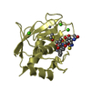
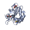
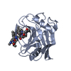
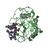
| Title | Structural and biochemical characterization of a cytosolic wheat cyclophilin TaCypA-1 | ||||||
 Components Components |
| ||||||
 Keywords Keywords | ISOMERASE/INHIBITOR /  ISOMERASE / ISOMERASE-INHIBITOR complex ISOMERASE / ISOMERASE-INHIBITOR complex | ||||||
| Function / homology |  Function and homology informationcyclosporin A binding / peptidylprolyl isomerase / peptidyl-prolyl cis-trans isomerase activity / protein folding / cytoplasm Function and homology informationcyclosporin A binding / peptidylprolyl isomerase / peptidyl-prolyl cis-trans isomerase activity / protein folding / cytoplasmSimilarity search - Function | ||||||
| Biological species |  Triticum aestivum (bread wheat) Triticum aestivum (bread wheat) Tolypocladium inflatum (fungus) Tolypocladium inflatum (fungus) | ||||||
| Method | X-RAY DIFFRACTION / SYNCHROTRON / MOLECULAR REPLACEMENT / Resolution: 1.2 Å | ||||||
 Authors Authors | Sekhon, S.S. / Jeong, D.G. / Woo, E.J. / Singh, P. / Pareek, A. / Yoon, T.-S. | ||||||
 Citation Citation | Journal: Acta Crystallogr.,Sect.D / Year: 2013 Title: Structural and biochemical characterization of the cytosolic wheat cyclophilin TaCypA-1. Authors: Sekhon, S.S. / Kaur, H. / Dutta, T. / Singh, K. / Kumari, S. / Kang, S. / Park, S.G. / Park, B.C. / Jeong, D.G. / Pareek, A. / Woo, E.J. / Singh, P. / Yoon, T.S. | ||||||
| History |
|
- Structure visualization
| Structure viewer | Molecule: MolmilJmol/JSmol |
|---|
- Downloads & links
-Download
| PDBx/mmCIF format | 4hy7.cif.gz | 53.6 KB | Display | PDBx/mmCIF format |
|---|---|---|---|---|
| PDB format | pdb4hy7.ent.gz | 36.2 KB | Display | PDB format |
| PDBx/mmJSON format | 4hy7.json.gz | Tree view | PDBx/mmJSON format | |
| Others |  Other downloads Other downloads |
-Validation report
| Arichive directory | https://data.pdbj.org/pub/pdb/validation_reports/hy/4hy7ftp://data.pdbj.org/pub/pdb/validation_reports/hy/4hy7 | HTTPS FTP |
|---|
-Related structure data
| Related structure data |  4e1qSC S: Starting model for refinement C: citing same article ( |
|---|---|
| Similar structure data |


-Links
 PDBj
PDBj

- Assembly
| Deposited unit | 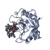
| ||||||||
|---|---|---|---|---|---|---|---|---|---|
| 1 |
| ||||||||
| Unit cell |
|
-Components
| #1: Protein | Prolyl isomerase Mass: 18416.098 Da / Num. of mol.: 1 Source method: isolated from a genetically manipulated source Source: (gene. exp.) Triticum aestivum (bread wheat) / Gene: CyP3, CyP1 / Production host:  Escherichia coli (E. coli) / References: UniProt: Q93W25, peptidylprolyl isomerase Escherichia coli (E. coli) / References: UniProt: Q93W25, peptidylprolyl isomerase |
|---|---|
| #2: Protein/peptide | Ciclosporin / Ciclosporin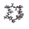  Type: Cyclic peptide / Class: ImmunosuppressantImmunosuppressive drug / Mass: 1220.625 Da / Num. of mol.: 1 / Source method: isolated from a natural source Type: Cyclic peptide / Class: ImmunosuppressantImmunosuppressive drug / Mass: 1220.625 Da / Num. of mol.: 1 / Source method: isolated from a natural sourceDetails: CYCLOSPORIN IS A CYCLIC UNDECAPEPTIDE. CYCLIZATION IS ACHIEVED BY LINKING THE N- AND THE C- TERMINI. Source: (natural) Tolypocladium inflatum (fungus) / References: NOR: NOR00033, Cyclosporin A |
| #3: Water | ChemComp-HOH / Water Mass: 18.015 Da / Num. of mol.: 158 / Source method: isolated from a natural source / Formula: H2O Mass: 18.015 Da / Num. of mol.: 158 / Source method: isolated from a natural source / Formula: H2O |
| Compound details | CYCLOSPORI |
-Experimental details
-Experiment
| Experiment | Method: X-RAY DIFFRACTION / Number of used crystals: 1 |
|---|
- Sample preparation
| Crystal | Density Matthews: 1.9 Å3/Da / Density % sol: 35.27 % |
|---|---|
| Crystal grow | Temperature: 291 K / Method: vapor diffusion, sitting drop / pH: 7.5 Details: 20% PEG 8000, 0.1M HEPES pH 7.5, VAPOR DIFFUSION, SITTING DROP, temperature 291K |
-Data collection
| Diffraction | Mean temperature: 197 K | ||||||||||||
|---|---|---|---|---|---|---|---|---|---|---|---|---|---|
| Diffraction source | Source: SYNCHROTRON / Wavelength: 0.9733 Å | ||||||||||||
| Detector | Type: ADSC QUANTUM 270 / Detector: CCD / Date: Oct 31, 2012 | ||||||||||||
| Radiation | Protocol: SINGLE WAVELENGTH / Monochromatic (M) / Laue (L): M / Scattering type: x-ray | ||||||||||||
| Radiation wavelength | Wavelength: 0.9733 Å / Relative weight: 1 | ||||||||||||
| Reflection | Resolution: 1.2→50 Å / Num. all: 47120 / Num. obs: 47120 / % possible obs: 99 % / Observed criterion σ(F): 0 / Observed criterion σ(I): -3 | ||||||||||||
| Reflection shell |
|
- Processing
| Software |
| |||||||||||||||||||||||||||||||||||||||||||||
|---|---|---|---|---|---|---|---|---|---|---|---|---|---|---|---|---|---|---|---|---|---|---|---|---|---|---|---|---|---|---|---|---|---|---|---|---|---|---|---|---|---|---|---|---|---|---|
| Refinement | Method to determine structure: MOLECULAR REPLACEMENT Starting model: 4E1Q Resolution: 1.2→27.35 Å / Cor.coef. Fo:Fc: 0.952 / Cor.coef. Fo:Fc free: 0.942 / SU B: 0.565 / SU ML: 0.027 / Cross valid method: THROUGHOUT / ESU R: 0.045 / ESU R Free: 0.046 / Stereochemistry target values: MAXIMUM LIKELIHOOD / Details: HYDROGENS HAVE BEEN USED IF PRESENT IN THE INPUT
| |||||||||||||||||||||||||||||||||||||||||||||
| Solvent computation | Ion probe radii: 0.8 Å / Shrinkage radii: 0.8 Å / VDW probe radii: 1.2 Å / Solvent model: MASK | |||||||||||||||||||||||||||||||||||||||||||||
| Displacement parameters | Biso mean: 11.143 Å2
| |||||||||||||||||||||||||||||||||||||||||||||
| Refinement step | Cycle: LAST / Resolution: 1.2→27.35 Å
| |||||||||||||||||||||||||||||||||||||||||||||
| Refine LS restraints |
| |||||||||||||||||||||||||||||||||||||||||||||
| LS refinement shell | Resolution: 1.2→1.231 Å / Total num. of bins used: 20
|
