 Movie
Movie Controller

[English] 日本語
 Yorodumi
- PDB-4hjt: Kinetic stabilization of transthyretin through covalent modificat... -
+ Open data

- Basic information
| Entry | Database: PDB / ID: 4hjt | ||||||
|---|---|---|---|---|---|---|---|
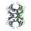
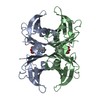
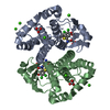
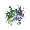
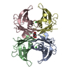
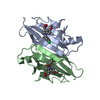
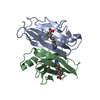
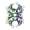
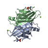
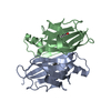
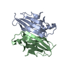
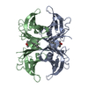
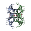
| Title | Kinetic stabilization of transthyretin through covalent modification of K15 by (E)-N-(4-(4-hydroxy-3,5-dimethylstyryl)phenyl)propionamide | ||||||
 Components Components | Transthyretin | ||||||
 Keywords Keywords | HORMONE BINDING PROTEIN/INHIBITOR / HORMONE BINDING PROTEIN / Thyroxine / retinol binding protein / HORMONE BINDING PROTEIN-INHIBITOR complex | ||||||
| Function / homology |  Function and homology information Function and homology informationRetinoid cycle disease events / The canonical retinoid cycle in rods (twilight vision) / thyroid hormone binding / purine nucleobase metabolic process / Non-integrin membrane-ECM interactions / Retinoid metabolism and transport / hormone activity / azurophil granule lumen / Amyloid fiber formation / Neutrophil degranulation ...Retinoid cycle disease events / The canonical retinoid cycle in rods (twilight vision) / thyroid hormone binding / purine nucleobase metabolic process / Non-integrin membrane-ECM interactions / Retinoid metabolism and transport / hormone activity / azurophil granule lumen / Amyloid fiber formation / Neutrophil degranulation / extracellular space / extracellular exosome / extracellular region / identical protein bindingSimilarity search - Function | ||||||
| Biological species |  Homo sapiens (human) Homo sapiens (human) | ||||||
| Method | X-RAY DIFFRACTION / SYNCHROTRON / MOLECULAR REPLACEMENT / Resolution: 1.45 Å | ||||||
 Authors Authors | Connelly, S. / Wilson, I.A. | ||||||
 Citation Citation | Journal: J.Am.Chem.Soc. / Year: 2013 Title: Stilbene vinyl sulfonamides as fluorogenic sensors of and traceless covalent kinetic stabilizers of transthyretin that prevent amyloidogenesis. Authors: Suh, E.H. / Liu, Y. / Connelly, S. / Genereux, J.C. / Wilson, I.A. / Kelly, J.W. | ||||||
| History |
|
- Structure visualization
| Structure viewer | Molecule: MolmilJmol/JSmol |
|---|
- Downloads & links
-Download
| PDBx/mmCIF format | 4hjt.cif.gz | 114.5 KB | Display | PDBx/mmCIF format |
|---|---|---|---|---|
| PDB format | pdb4hjt.ent.gz | 89.7 KB | Display | PDB format |
| PDBx/mmJSON format | 4hjt.json.gz | Tree view | PDBx/mmJSON format | |
| Others |  Other downloads Other downloads |
-Validation report
| Arichive directory | https://data.pdbj.org/pub/pdb/validation_reports/hj/4hjtftp://data.pdbj.org/pub/pdb/validation_reports/hj/4hjt | HTTPS FTP |
|---|
-Related structure data
| Related structure data | 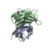 4hjsC  4hjuC 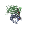 2fbrS C: citing same article ( S: Starting model for refinement |
|---|---|
| Similar structure data |


-Links
 PDBj
PDBj


- Assembly
| Deposited unit | 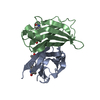
| |||||||||||||||||||||||||||
|---|---|---|---|---|---|---|---|---|---|---|---|---|---|---|---|---|---|---|---|---|---|---|---|---|---|---|---|---|
| 1 | 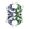
| |||||||||||||||||||||||||||
| Unit cell |
| |||||||||||||||||||||||||||
| Components on special symmetry positions |
|
-Components
| #1: Protein | / ATTR / Prealbumin / TBPA Mass: 13777.360 Da / Num. of mol.: 2 Source method: isolated from a genetically manipulated source Source: (gene. exp.) Homo sapiens (human) / Gene: PALB, TTR / Plasmid: pmmHA / Production host:  Escherichia coli (E. coli) / Strain (production host): Epicurean Gold / References: UniProt: P02766 Escherichia coli (E. coli) / Strain (production host): Epicurean Gold / References: UniProt: P02766#2: Chemical |   Mass: 295.376 Da / Num. of mol.: 2 / Source method: obtained synthetically / Formula: C19H21NO2 Mass: 295.376 Da / Num. of mol.: 2 / Source method: obtained synthetically / Formula: C19H21NO2#3: Water | ChemComp-HOH / | Water Mass: 18.015 Da / Num. of mol.: 82 / Source method: isolated from a natural source / Formula: H2O Mass: 18.015 Da / Num. of mol.: 82 / Source method: isolated from a natural source / Formula: H2O |
|---|
-Experimental details
-Experiment
| Experiment | Method: X-RAY DIFFRACTION / Number of used crystals: 1 |
|---|
- Sample preparation
| Crystal | Density Matthews: 2.13 Å3/Da / Density % sol: 42.22 % |
|---|---|
| Crystal grow | Temperature: 298 K / Method: vapor diffusion, sitting drop / pH: 5.5 Details: THE WT-TTR WAS CONCENTRATED TO 4 MG/ML IN 10 MM NAPI, 100 MM KCL, AT PH 7.6 AND CO-CRYSTALLIZED AT ROOM TEMPERATURE WITH INHIBITORS USING THE VAPOR-DIFFUSION SITTING DROP METHOD, CRYSTALS ...Details: THE WT-TTR WAS CONCENTRATED TO 4 MG/ML IN 10 MM NAPI, 100 MM KCL, AT PH 7.6 AND CO-CRYSTALLIZED AT ROOM TEMPERATURE WITH INHIBITORS USING THE VAPOR-DIFFUSION SITTING DROP METHOD, CRYSTALS WERE GROWN FROM 1.395 M SODIUM CITRATE, 3.5% V/V GLYCEROL AT PH 5.5. THE CRYSTALS WERE FROZEN USING A CRYO-PROTECTANT SOLUTION OF 1.395 M SODIUM CITRATE, PH 5.5, CONTAINING 10% V/V GLYCEROL, VAPOR DIFFUSION, SITTING DROP, temperature 298K |
-Data collection
| Diffraction source | Source: SYNCHROTRON / Site: SSRL  / Beamline: BL11-1 / Wavelength: 0.9795 Å / Beamline: BL11-1 / Wavelength: 0.9795 Å |
|---|---|
| Detector | Type: PSI PILATUS 6M / Detector: PIXEL / Date: Apr 6, 2012 / Details: Rh coated flat mirror |
| Radiation | Monochromator: Side scattering bent cube-root I-beam single crystal; asymmetric cut 4.965 degs Protocol: SINGLE WAVELENGTH / Monochromatic (M) / Laue (L): M / Scattering type: x-ray |
| Radiation wavelength | Wavelength: 0.9795 Å / Relative weight: 1 |
| Reflection | Resolution: 1.45→85.16 Å / Num. obs: 42818 / % possible obs: 99.9 % / Redundancy: 8.2 % / Biso Wilson estimate: 20.4 Å2 / Rsym value: 0.044 / Net I/σ(I): 55.9 |
| Reflection shell | Resolution: 1.45→1.5 Å / Redundancy: 8.1 % / Mean I/σ(I) obs: 3.7 / Num. unique all: 4221 / Rsym value: 0.519 / % possible all: 100 |
- Processing
| Software |
| ||||||||||||||||||||||||||||||||||||||||||||||||||||||||||||||||||||||||||||||||||||||||||
|---|---|---|---|---|---|---|---|---|---|---|---|---|---|---|---|---|---|---|---|---|---|---|---|---|---|---|---|---|---|---|---|---|---|---|---|---|---|---|---|---|---|---|---|---|---|---|---|---|---|---|---|---|---|---|---|---|---|---|---|---|---|---|---|---|---|---|---|---|---|---|---|---|---|---|---|---|---|---|---|---|---|---|---|---|---|---|---|---|---|---|---|
| Refinement | Method to determine structure: MOLECULAR REPLACEMENT Starting model: 2FBR Resolution: 1.45→85.16 Å / Cor.coef. Fo:Fc: 0.97 / Cor.coef. Fo:Fc free: 0.963 / WRfactor Rfree: 0.1922 / WRfactor Rwork: 0.1698 / Occupancy max: 1 / Occupancy min: 0.25 / FOM work R set: 0.9088 / SU B: 1.802 / SU ML: 0.032 / SU R Cruickshank DPI: 0.0772 / SU Rfree: 0.0634 / Cross valid method: THROUGHOUT / σ(F): 0 / ESU R: 0.077 / ESU R Free: 0.063 / Stereochemistry target values: MAXIMUM LIKELIHOOD Details: HYDROGENS HAVE BEEN ADDED IN THE RIDING POSITIONS U VALUES : REFINED INDIVIDUALLY
| ||||||||||||||||||||||||||||||||||||||||||||||||||||||||||||||||||||||||||||||||||||||||||
| Solvent computation | Ion probe radii: 0.8 Å / Shrinkage radii: 0.8 Å / VDW probe radii: 1.4 Å / Solvent model: MASK | ||||||||||||||||||||||||||||||||||||||||||||||||||||||||||||||||||||||||||||||||||||||||||
| Displacement parameters | Biso max: 156.33 Å2 / Biso mean: 23.2489 Å2 / Biso min: 10.25 Å2
| ||||||||||||||||||||||||||||||||||||||||||||||||||||||||||||||||||||||||||||||||||||||||||
| Refinement step | Cycle: LAST / Resolution: 1.45→85.16 Å
| ||||||||||||||||||||||||||||||||||||||||||||||||||||||||||||||||||||||||||||||||||||||||||
| Refine LS restraints |
| ||||||||||||||||||||||||||||||||||||||||||||||||||||||||||||||||||||||||||||||||||||||||||
| LS refinement shell | Resolution: 1.45→1.488 Å / Total num. of bins used: 20
|
