 Movie
Movie Controller

[English] 日本語
 Yorodumi
- PDB-4hjc: Crystal structure of glycoprotein C from Rift Valley Fever Virus ... -
+ Open data

- Basic information
| Entry | Database: PDB / ID: 4hjc | ||||||
|---|---|---|---|---|---|---|---|
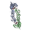
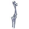
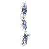
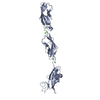
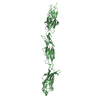
| Title | Crystal structure of glycoprotein C from Rift Valley Fever Virus (non-glycosylated) | ||||||
 Components Components | ENVELOPE GLYCOPROTEIN | ||||||
 Keywords Keywords |  VIRAL PROTEIN / virus entry / class II fusion protein / membrane fusion / viral membrane VIRAL PROTEIN / virus entry / class II fusion protein / membrane fusion / viral membrane | ||||||
| Function / homology |  Function and homology information Function and homology informationhost cell mitochondrial outer membrane / : / host cell Golgi membrane / : / entry receptor-mediated virion attachment to host cell / host cell endoplasmic reticulum membrane / symbiont entry into host cell / fusion of virus membrane with host endosome membrane / virion membrane / membraneSimilarity search - Function | ||||||
| Biological species |   Rift Valley fever virus Rift Valley fever virus | ||||||
| Method | X-RAY DIFFRACTION / SYNCHROTRON / MOLECULAR REPLACEMENT / molecular replacement / Resolution: 4.15 Å | ||||||
 Authors Authors | Dessau, M. / Modis, Y. | ||||||
 Citation Citation | Journal: Proc.Natl.Acad.Sci.USA / Year: 2013 Title: Crystal structure of glycoprotein C from Rift Valley fever virus. Authors: Dessau, M. / Modis, Y. | ||||||
| History |
|
- Structure visualization
| Structure viewer | Molecule: MolmilJmol/JSmol |
|---|
- Downloads & links
-Download
| PDBx/mmCIF format | 4hjc.cif.gz | 177 KB | Display | PDBx/mmCIF format |
|---|---|---|---|---|
| PDB format | pdb4hjc.ent.gz | 148.6 KB | Display | PDB format |
| PDBx/mmJSON format | 4hjc.json.gz | Tree view | PDBx/mmJSON format | |
| Others |  Other downloads Other downloads |
-Validation report
| Arichive directory | https://data.pdbj.org/pub/pdb/validation_reports/hj/4hjcftp://data.pdbj.org/pub/pdb/validation_reports/hj/4hjc | HTTPS FTP |
|---|
-Related structure data


-Links
 PDBj
PDBj- Assembly
| Deposited unit | 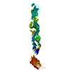
| ||||||||
|---|---|---|---|---|---|---|---|---|---|
| 1 |
| ||||||||
| Unit cell |
|
-Components
| #1: Protein | Mass: 46586.316 Da / Num. of mol.: 1 Source method: isolated from a genetically manipulated source Source: (gene. exp.) Rift Valley fever virus / Plasmid: pAc-gp67 / Production host: unidentified baculovirus / References: UniProt: A2T075, UniProt: P03518*PLUS |
|---|---|
| #2: Water | ChemComp-HOH / Water Mass: 18.015 Da / Num. of mol.: 5 / Source method: isolated from a natural source / Formula: H2O Mass: 18.015 Da / Num. of mol.: 5 / Source method: isolated from a natural source / Formula: H2O |
-Experimental details
-Experiment
| Experiment | Method: X-RAY DIFFRACTION / Number of used crystals: 1 |
|---|
- Sample preparation
| Crystal | Density Matthews: 3.46 Å3/Da / Density % sol: 64.42 % |
|---|---|
| Crystal grow | Temperature: 289 K / Method: vapor diffusion, hanging drop / pH: 6.2 Details: 12% PEG 5000 MME, 0.1 M MES, O.1 M Ammonium sulfate, 5% glycerol, 1.8 mM UDM, pH 6.2, VAPOR DIFFUSION, HANGING DROP, temperature 289K |
-Data collection
| Diffraction | Mean temperature: 100 K | |||||||||||||||||||||||||||||||||||||||||||||||||||||||||||||||||||||||||||||||||||||||||||||||||||||||||||||||||||||||||||||||||||||||||||||||||||
|---|---|---|---|---|---|---|---|---|---|---|---|---|---|---|---|---|---|---|---|---|---|---|---|---|---|---|---|---|---|---|---|---|---|---|---|---|---|---|---|---|---|---|---|---|---|---|---|---|---|---|---|---|---|---|---|---|---|---|---|---|---|---|---|---|---|---|---|---|---|---|---|---|---|---|---|---|---|---|---|---|---|---|---|---|---|---|---|---|---|---|---|---|---|---|---|---|---|---|---|---|---|---|---|---|---|---|---|---|---|---|---|---|---|---|---|---|---|---|---|---|---|---|---|---|---|---|---|---|---|---|---|---|---|---|---|---|---|---|---|---|---|---|---|---|---|---|---|---|
| Diffraction source | Source: SYNCHROTRON / Site: NSLS  / Beamline: X25 / Wavelength: 1.1 Å / Beamline: X25 / Wavelength: 1.1 Å | |||||||||||||||||||||||||||||||||||||||||||||||||||||||||||||||||||||||||||||||||||||||||||||||||||||||||||||||||||||||||||||||||||||||||||||||||||
| Detector | Type: PSI PILATUS 6M / Detector: PIXEL / Date: Jul 18, 2012 | |||||||||||||||||||||||||||||||||||||||||||||||||||||||||||||||||||||||||||||||||||||||||||||||||||||||||||||||||||||||||||||||||||||||||||||||||||
| Radiation | Protocol: SINGLE WAVELENGTH / Monochromatic (M) / Laue (L): M / Scattering type: x-ray | |||||||||||||||||||||||||||||||||||||||||||||||||||||||||||||||||||||||||||||||||||||||||||||||||||||||||||||||||||||||||||||||||||||||||||||||||||
| Radiation wavelength | Wavelength: 1.1 Å / Relative weight: 1 | |||||||||||||||||||||||||||||||||||||||||||||||||||||||||||||||||||||||||||||||||||||||||||||||||||||||||||||||||||||||||||||||||||||||||||||||||||
| Reflection | Resolution: 4.15→40 Å / Num. all: 5449 / Num. obs: 5438 / % possible obs: 99.8 % / Observed criterion σ(F): 2.8 / Observed criterion σ(I): 1.4 / Redundancy: 12.6 % / Rmerge(I) obs: 0.075 / Χ2: 0.714 / Net I/σ(I): 5 | |||||||||||||||||||||||||||||||||||||||||||||||||||||||||||||||||||||||||||||||||||||||||||||||||||||||||||||||||||||||||||||||||||||||||||||||||||
| Reflection shell |
|
-Phasing
| Phasing | Method: molecular replacement |
|---|
- Processing
| Software |
| ||||||||||||||||||||||||||||||||||||||||
|---|---|---|---|---|---|---|---|---|---|---|---|---|---|---|---|---|---|---|---|---|---|---|---|---|---|---|---|---|---|---|---|---|---|---|---|---|---|---|---|---|---|
| Refinement | Method to determine structure: MOLECULAR REPLACEMENT / Resolution: 4.15→19.988 Å / Occupancy max: 1 / Occupancy min: 1 / FOM work R set: 0.6472 / SU ML: 0.55 / σ(F): 1.39 / Phase error: 38.31 / Stereochemistry target values: ML
| ||||||||||||||||||||||||||||||||||||||||
| Solvent computation | Shrinkage radii: 0.9 Å / VDW probe radii: 1.11 Å / Solvent model: FLAT BULK SOLVENT MODEL | ||||||||||||||||||||||||||||||||||||||||
| Displacement parameters | Biso max: 465.24 Å2 / Biso mean: 173.1681 Å2 / Biso min: 20 Å2 | ||||||||||||||||||||||||||||||||||||||||
| Refinement step | Cycle: LAST / Resolution: 4.15→19.988 Å
| ||||||||||||||||||||||||||||||||||||||||
| Refine LS restraints |
| ||||||||||||||||||||||||||||||||||||||||
| LS refinement shell | Refine-ID: X-RAY DIFFRACTION / Total num. of bins used: 2 / % reflection obs: 100 %
| ||||||||||||||||||||||||||||||||||||||||
| Refinement TLS params. | Method: refined / Origin x: 155.7479 Å / Origin y: 150.1085 Å / Origin z: 33.3301 Å
| ||||||||||||||||||||||||||||||||||||||||
| Refinement TLS group |
|
