 Movie
Movie Controller

[English] 日本語
 Yorodumi
- PDB-4hic: Crystal structure of the potential transfer protein TraK from Gra... -
+ Open data

- Basic information
| Entry | Database: PDB / ID: 4hic | ||||||
|---|---|---|---|---|---|---|---|
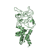
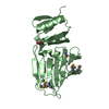
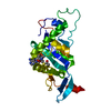
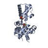
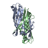
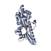
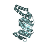
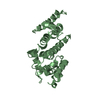
| Title | Crystal structure of the potential transfer protein TraK from Gram-positive conjugative plasmid pIP501 | ||||||
 Components Components | TraK | ||||||
 Keywords Keywords | UNKNOWN FUNCTION / Gram-positive / type-IV secretion / pIP501 / anti-parallel beta-sheets | ||||||
| Function / homology | membrane => GO:0016020 / Uncharacterized protein Function and homology information Function and homology information | ||||||
| Biological species |   Enterococcus faecalis (bacteria) Enterococcus faecalis (bacteria) | ||||||
| Method | X-RAY DIFFRACTION / SYNCHROTRON / MOLECULAR REPLACEMENT / molecular replacement / Resolution: 3.001 Å | ||||||
 Authors Authors | Goessweiner-Mohr, N. / Keller, W. | ||||||
 Citation Citation | Journal: Acta Crystallogr.,Sect.D / Year: 2014 Title: The type IV secretion protein TraK from the Enterococcus conjugative plasmid pIP501 exhibits a novel fold Authors: Goessweiner-Mohr, N. / Fercher, C. / Arends, K. / Birner-Gruenberger, R. / Laverde-Gomez, D. / Huebner, J. / Grohmann, E. / Keller, W. | ||||||
| History |
|
- Structure visualization
| Structure viewer | Molecule: MolmilJmol/JSmol |
|---|
- Downloads & links
-Download
| PDBx/mmCIF format | 4hic.cif.gz | 94.5 KB | Display | PDBx/mmCIF format |
|---|---|---|---|---|
| PDB format | pdb4hic.ent.gz | 71.9 KB | Display | PDB format |
| PDBx/mmJSON format | 4hic.json.gz | Tree view | PDBx/mmJSON format | |
| Others |  Other downloads Other downloads |
-Validation report
| Arichive directory | https://data.pdbj.org/pub/pdb/validation_reports/hi/4hicftp://data.pdbj.org/pub/pdb/validation_reports/hi/4hic | HTTPS FTP |
|---|
-Related structure data
| Similar structure data |
|---|
-Links
 PDBj
PDBj- Assembly
| Deposited unit | 
| ||||||||
|---|---|---|---|---|---|---|---|---|---|
| 1 | 
| ||||||||
| 2 | 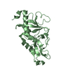
| ||||||||
| Unit cell |
|
-Components
| #1: Protein | Mass: 30643.514 Da / Num. of mol.: 2 Source method: isolated from a genetically manipulated source Source: (gene. exp.) Enterococcus faecalis (bacteria) / Gene: ORF11 from conjugative plasmid pIP501 / Plasmid: pQTEV / Production host: Escherichia coli (E. coli) / Strain (production host): BL21 (DE3) / References: UniProt: Q8L1C9 |
|---|
-Experimental details
-Experiment
| Experiment | Method: X-RAY DIFFRACTION / Number of used crystals: 1 |
|---|
- Sample preparation
| Crystal | Density Matthews: 3.2 Å3/Da / Density % sol: 61.52 % |
|---|---|
| Crystal grow | Temperature: 298 K / Method: vapor diffusion: microbatch under oil / pH: 6.5 Details: 1:1 setup of purification buffer with Morpheus screen condition 52 (12.5 % PEG 1000, 12.5 % PEG 3350, 12.5 % MPD, ethylene glycols mix, 0.1 M MES/Imidazol); final protein concentration: 6.45 ...Details: 1:1 setup of purification buffer with Morpheus screen condition 52 (12.5 % PEG 1000, 12.5 % PEG 3350, 12.5 % MPD, ethylene glycols mix, 0.1 M MES/Imidazol); final protein concentration: 6.45 mg/ml, pH 6.5, vapor diffusion: microbatch under oil, temperature 298K |
-Data collection
| Diffraction | Mean temperature: 100 K | ||||||||||||||||||||||||||||||||||||||||||||||||||||||||||||||||||||||||||||||||||||||||
|---|---|---|---|---|---|---|---|---|---|---|---|---|---|---|---|---|---|---|---|---|---|---|---|---|---|---|---|---|---|---|---|---|---|---|---|---|---|---|---|---|---|---|---|---|---|---|---|---|---|---|---|---|---|---|---|---|---|---|---|---|---|---|---|---|---|---|---|---|---|---|---|---|---|---|---|---|---|---|---|---|---|---|---|---|---|---|---|---|---|
| Diffraction source | Source: SYNCHROTRON / Site: SLS  / Beamline: X06DA / Wavelength: 1 Å / Beamline: X06DA / Wavelength: 1 Å | ||||||||||||||||||||||||||||||||||||||||||||||||||||||||||||||||||||||||||||||||||||||||
| Detector | Type: MARMOSAIC 225 mm CCD / Detector: CCD / Date: May 30, 2010 Details: toroidal mirror (M2) to vertically and horizontally focus the beam at the sample position (with 2:1 horizontal demagnification) | ||||||||||||||||||||||||||||||||||||||||||||||||||||||||||||||||||||||||||||||||||||||||
| Radiation | Monochromator: BARTELS MONOCROMATOR / Protocol: Native / Scattering type: x-ray | ||||||||||||||||||||||||||||||||||||||||||||||||||||||||||||||||||||||||||||||||||||||||
| Radiation wavelength | Wavelength: 1 Å / Relative weight: 1 | ||||||||||||||||||||||||||||||||||||||||||||||||||||||||||||||||||||||||||||||||||||||||
| Reflection twin | Operator: -h,k,-l / Fraction: 0.24 | ||||||||||||||||||||||||||||||||||||||||||||||||||||||||||||||||||||||||||||||||||||||||
| Reflection | Resolution: 3.001→82.835 Å / Num. all: 15465 / Num. obs: 15465 / % possible obs: 99.9 % / Observed criterion σ(F): 0 / Observed criterion σ(I): 0 / Redundancy: 7.6 % / Rsym value: 0.094 / Net I/σ(I): 12.3 | ||||||||||||||||||||||||||||||||||||||||||||||||||||||||||||||||||||||||||||||||||||||||
| Reflection shell | Diffraction-ID: 1
|
-Phasing
| Phasing | Method: molecular replacement | |||||||||
|---|---|---|---|---|---|---|---|---|---|---|
| Phasing MR |
|
- Processing
| Software |
| ||||||||||||||||||||||||||||||||||||
|---|---|---|---|---|---|---|---|---|---|---|---|---|---|---|---|---|---|---|---|---|---|---|---|---|---|---|---|---|---|---|---|---|---|---|---|---|---|
| Refinement | Method to determine structure: MOLECULAR REPLACEMENT / Resolution: 3.001→40.319 Å / Occupancy max: 1 / Occupancy min: 1 / FOM work R set: 0.7381 / σ(F): 0 / Phase error: 33.08 / Stereochemistry target values: TWIN_LSQ_F
| ||||||||||||||||||||||||||||||||||||
| Solvent computation | Shrinkage radii: 0.9 Å / VDW probe radii: 1.11 Å / Solvent model: FLAT BULK SOLVENT MODEL | ||||||||||||||||||||||||||||||||||||
| Displacement parameters | Biso max: 126.11 Å2 / Biso mean: 59.7934 Å2 / Biso min: 32.07 Å2 | ||||||||||||||||||||||||||||||||||||
| Refinement step | Cycle: LAST / Resolution: 3.001→40.319 Å
| ||||||||||||||||||||||||||||||||||||
| Refine LS restraints |
| ||||||||||||||||||||||||||||||||||||
| LS refinement shell | Refine-ID: X-RAY DIFFRACTION / Total num. of bins used: 5 / % reflection obs: 95 %
|
