 Movie
Movie Controller


+ Open data

- Basic information
| Entry | Database: PDB / ID: 4hh8 | ||||||
|---|---|---|---|---|---|---|---|
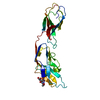
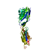
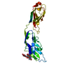
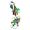
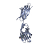
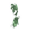
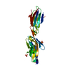
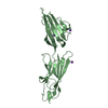
| Title | Crystal structure of bovine butyrophilin | ||||||
 Components Components | Butyrophilin subfamily 1 member A1 | ||||||
 Keywords Keywords |  PROTEIN BINDING / Immunoglobulin Fold / milk fat globule PROTEIN BINDING / Immunoglobulin Fold / milk fat globule | ||||||
| Function / homology |  Function and homology informationregulation of immune response / regulation of cytokine production / T cell receptor signaling pathway / membrane => GO:0016020 / external side of plasma membrane / signaling receptor binding Function and homology informationregulation of immune response / regulation of cytokine production / T cell receptor signaling pathway / membrane => GO:0016020 / external side of plasma membrane / signaling receptor bindingSimilarity search - Function | ||||||
| Biological species |  Bos taurus (cattle) Bos taurus (cattle) | ||||||
| Method | X-RAY DIFFRACTION / SYNCHROTRON / MAD / Resolution: 2.3 Å | ||||||
 Authors Authors | Eichinger, A. / Skerra, A. | ||||||
 Citation Citation | Journal: To be Published Title: The extracellular region of bovine butyrophilin exhibits high structural similarity to human myelin oligodendrocyte glycoprotein Authors: Eichinger, A. / Neumaier, I. / Skerra, A. | ||||||
| History |
|
- Structure visualization
| Structure viewer | Molecule: MolmilJmol/JSmol |
|---|
- Downloads & links
-Download
| PDBx/mmCIF format | 4hh8.cif.gz | 53.2 KB | Display | PDBx/mmCIF format |
|---|---|---|---|---|
| PDB format | pdb4hh8.ent.gz | 40.6 KB | Display | PDB format |
| PDBx/mmJSON format | 4hh8.json.gz | Tree view | PDBx/mmJSON format | |
| Others |  Other downloads Other downloads |
-Validation report
| Arichive directory | https://data.pdbj.org/pub/pdb/validation_reports/hh/4hh8ftp://data.pdbj.org/pub/pdb/validation_reports/hh/4hh8 | HTTPS FTP |
|---|
-Related structure data
| Similar structure data |
|---|

-Links
 PDBj
PDBj


- Assembly
| Deposited unit | 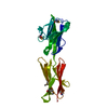
| ||||||||
|---|---|---|---|---|---|---|---|---|---|
| 1 |
| ||||||||
| Unit cell |
|
-Components
| #1: Protein | Mass: 25279.395 Da / Num. of mol.: 1 / Mutation: C193A Source method: isolated from a genetically manipulated source Source: (gene. exp.) Bos taurus (cattle) / Gene: BTN, BTN1A1 / Production host:  Escherichia coli (E. coli) / Strain (production host): JM83 / References: UniProt: P18892 Escherichia coli (E. coli) / Strain (production host): JM83 / References: UniProt: P18892 |
|---|---|
| #2: Water | ChemComp-HOH / Water Mass: 18.015 Da / Num. of mol.: 55 / Source method: isolated from a natural source / Formula: H2O Mass: 18.015 Da / Num. of mol.: 55 / Source method: isolated from a natural source / Formula: H2O |
| Sequence details | THE DIFFERENCES BETWEEN THE SEQUENCE OF THE SOLVED STRUCTURE AND THE DATABASE ENTRY P18892 ARISE ...THE DIFFERENCE |
-Experimental details
-Experiment
| Experiment | Method: X-RAY DIFFRACTION / Number of used crystals: 1 |
|---|
- Sample preparation
| Crystal | Density Matthews: 3.24 Å3/Da / Density % sol: 62.03 % |
|---|---|
| Crystal grow | Temperature: 293 K / Method: vapor diffusion, hanging drop / pH: 7.4 Details: 1.5 M potassium phosphate, pH 7.4, VAPOR DIFFUSION, HANGING DROP, temperature 293K |
-Data collection
| Diffraction | Mean temperature: 100 K | ||||||||||||||||||||||||||||||||||||||||||||||||||||||||||||||||||||||||||||||||||||||||
|---|---|---|---|---|---|---|---|---|---|---|---|---|---|---|---|---|---|---|---|---|---|---|---|---|---|---|---|---|---|---|---|---|---|---|---|---|---|---|---|---|---|---|---|---|---|---|---|---|---|---|---|---|---|---|---|---|---|---|---|---|---|---|---|---|---|---|---|---|---|---|---|---|---|---|---|---|---|---|---|---|---|---|---|---|---|---|---|---|---|
| Diffraction source | Source: SYNCHROTRON / Site: BESSY  / Beamline: 14.1 / Wavelength: 0.97973,0.97985,0.96863,0.99188 / Beamline: 14.1 / Wavelength: 0.97973,0.97985,0.96863,0.99188 | ||||||||||||||||||||||||||||||||||||||||||||||||||||||||||||||||||||||||||||||||||||||||
| Detector | Type: MARMOSAIC 225 mm CCD / Detector: CCD / Date: Dec 15, 2009 / Details: mirrors | ||||||||||||||||||||||||||||||||||||||||||||||||||||||||||||||||||||||||||||||||||||||||
| Radiation | Monochromator: Si 111 CRYSTAL / Protocol: MAD / Monochromatic (M) / Laue (L): M / Scattering type: x-ray | ||||||||||||||||||||||||||||||||||||||||||||||||||||||||||||||||||||||||||||||||||||||||
| Radiation wavelength |
| ||||||||||||||||||||||||||||||||||||||||||||||||||||||||||||||||||||||||||||||||||||||||
| Reflection | Redundancy: 3.9 % / Av σ(I) over netI: 14.1 / Number: 32994 / Rsym value: 0.033 / D res high: 2.8 Å / D res low: 59.188 Å / Num. obs: 8446 / % possible obs: 99.4 | ||||||||||||||||||||||||||||||||||||||||||||||||||||||||||||||||||||||||||||||||||||||||
| Diffraction reflection shell |
| ||||||||||||||||||||||||||||||||||||||||||||||||||||||||||||||||||||||||||||||||||||||||
| Reflection | Resolution: 2.3→59.264 Å / Num. all: 15058 / Num. obs: 15058 / % possible obs: 99.3 % / Observed criterion σ(F): 2 / Observed criterion σ(I): 2 / Redundancy: 7.1 % / Rsym value: 0.059 / Net I/σ(I): 16.7 | ||||||||||||||||||||||||||||||||||||||||||||||||||||||||||||||||||||||||||||||||||||||||
| Reflection shell | Diffraction-ID: 1
|
-Phasing
| Phasing | Method: MAD |
|---|
- Processing
| Software |
| |||||||||||||||||||||||||||||||||||||||||||||||||||||||||||||||||
|---|---|---|---|---|---|---|---|---|---|---|---|---|---|---|---|---|---|---|---|---|---|---|---|---|---|---|---|---|---|---|---|---|---|---|---|---|---|---|---|---|---|---|---|---|---|---|---|---|---|---|---|---|---|---|---|---|---|---|---|---|---|---|---|---|---|---|
| Refinement | Method to determine structure: MAD / Resolution: 2.3→59.26 Å / Cor.coef. Fo:Fc: 0.935 / Cor.coef. Fo:Fc free: 0.907 / WRfactor Rfree: 0.2848 / WRfactor Rwork: 0.2323 / Occupancy max: 1 / Occupancy min: 1 / FOM work R set: 0.8146 / SU B: 5.777 / SU ML: 0.146 / SU R Cruickshank DPI: 0.2541 / SU Rfree: 0.2263 / Cross valid method: THROUGHOUT / σ(F): 2 / ESU R: 0.254 / ESU R Free: 0.226 / Stereochemistry target values: MAXIMUM LIKELIHOOD
| |||||||||||||||||||||||||||||||||||||||||||||||||||||||||||||||||
| Solvent computation | Ion probe radii: 0.8 Å / Shrinkage radii: 0.8 Å / VDW probe radii: 1.4 Å / Solvent model: MASK | |||||||||||||||||||||||||||||||||||||||||||||||||||||||||||||||||
| Displacement parameters | Biso max: 90.5 Å2 / Biso mean: 42.1194 Å2 / Biso min: 21.09 Å2
| |||||||||||||||||||||||||||||||||||||||||||||||||||||||||||||||||
| Refinement step | Cycle: LAST / Resolution: 2.3→59.26 Å
| |||||||||||||||||||||||||||||||||||||||||||||||||||||||||||||||||
| Refine LS restraints |
| |||||||||||||||||||||||||||||||||||||||||||||||||||||||||||||||||
| LS refinement shell | Resolution: 2.3→2.36 Å / Total num. of bins used: 20
|
