 Movie
Movie Controller


+ Open data

- Basic information
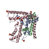
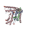
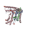
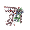
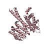
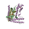
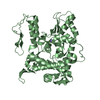
| Entry | Database: PDB / ID: 4h9n | ||||||
|---|---|---|---|---|---|---|---|
| Title | Complex structure 1 of DAXX/H3.3(sub5)/H4 | ||||||
 Components Components |
| ||||||
 Keywords Keywords | DNA BINDING PROTEIN/APOPTOSIS / histone chaperone / DNA BINDING PROTEIN-APOPTOSIS complex | ||||||
| Function / homology |  Function and homology information Function and homology informationcellular response to diamide / Defective Inhibition of DNA Recombination at Telomere Due to DAXX Mutations / Defective Inhibition of DNA Recombination at Telomere Due to ATRX Mutations / SUMO-modified protein reader activity / neuron intrinsic apoptotic signaling pathway in response to oxidative stress / cellular response to sodium arsenite / negative regulation of chromosome condensation /  Barr body / regulation of centromere complex assembly / muscle cell differentiation ...cellular response to diamide / Defective Inhibition of DNA Recombination at Telomere Due to DAXX Mutations / Defective Inhibition of DNA Recombination at Telomere Due to ATRX Mutations / SUMO-modified protein reader activity / neuron intrinsic apoptotic signaling pathway in response to oxidative stress / cellular response to sodium arsenite / negative regulation of chromosome condensation / Barr body / regulation of centromere complex assembly / muscle cell differentiation / pericentric heterochromatin formation / inner kinetochore / transcription regulator inhibitor activity / oocyte maturation / nuclear androgen receptor binding / nucleus organization / protein kinase activator activity / androgen receptor signaling pathway / chromosome, centromeric region / regulation of protein ubiquitination / spermatid development / extrinsic apoptotic signaling pathway via death domain receptors / single fertilization / subtelomeric heterochromatin formation / negative regulation of megakaryocyte differentiation / positive regulation of protein kinase activity / RNA polymerase II core promoter sequence-specific DNA binding / cellular response to unfolded protein / nucleosomal DNA binding / protein localization to CENP-A containing chromatin / Replacement of protamines by nucleosomes in the male pronucleus / CENP-A containing nucleosome / Packaging Of Telomere Ends / Recognition and association of DNA glycosylase with site containing an affected purine / Cleavage of the damaged purine / JNK cascade / Deposition of new CENPA-containing nucleosomes at the centromere / cellular response to copper ion / Recognition and association of DNA glycosylase with site containing an affected pyrimidine / Cleavage of the damaged pyrimidine / Inhibition of DNA recombination at telomere / heat shock protein binding / Meiotic synapsis / cellular response to cadmium ion / telomere organization / embryo implantation / RNA Polymerase I Promoter Opening / Assembly of the ORC complex at the origin of replication / SUMOylation of chromatin organization proteins / DNA methylation / Condensation of Prophase Chromosomes / SUMOylation of transcription cofactors / ERCC6 (CSB) and EHMT2 (G9a) positively regulate rRNA expression / SIRT1 negatively regulates rRNA expression / Chromatin modifications during the maternal to zygotic transition (MZT) / HCMV Late Events / PRC2 methylates histones and DNA / Defective pyroptosis / molecular condensate scaffold activity / HDACs deacetylate histones / RNA Polymerase I Promoter Escape / Nonhomologous End-Joining (NHEJ) / Transcriptional regulation by small RNAs / Formation of the beta-catenin:TCF transactivating complex / RUNX1 regulates genes involved in megakaryocyte differentiation and platelet function / Activated PKN1 stimulates transcription of AR (androgen receptor) regulated genes KLK2 and KLK3 / NoRC negatively regulates rRNA expression / G2/M DNA damage checkpoint / B-WICH complex positively regulates rRNA expression / HDMs demethylate histones / multicellular organism growth / DNA Damage/Telomere Stress Induced Senescence / PKMTs methylate histone lysines / PML body / RMTs methylate histone arginines / Meiotic recombination / Pre-NOTCH Transcription and Translation / Activation of anterior HOX genes in hindbrain development during early embryogenesis / HCMV Early Events / osteoblast differentiation / Transcriptional regulation of granulopoiesis / structural constituent of chromatin / transcription corepressor activity / male gonad development / nucleosome / nucleosome assembly / Regulation of TP53 Degradation / p53 binding / Recruitment and ATM-mediated phosphorylation of repair and signaling proteins at DNA double strand breaks / RUNX1 regulates transcription of genes involved in differentiation of HSCs / chromatin organization / cellular response to heat / Factors involved in megakaryocyte development and platelet production / HATs acetylate histones / Processing of DNA double-strand break ends / histone binding / Senescence-Associated Secretory Phenotype (SASP) / positive regulation of cell growth / regulation of gene expression / regulation of apoptotic process Barr body / regulation of centromere complex assembly / muscle cell differentiation ...cellular response to diamide / Defective Inhibition of DNA Recombination at Telomere Due to DAXX Mutations / Defective Inhibition of DNA Recombination at Telomere Due to ATRX Mutations / SUMO-modified protein reader activity / neuron intrinsic apoptotic signaling pathway in response to oxidative stress / cellular response to sodium arsenite / negative regulation of chromosome condensation / Barr body / regulation of centromere complex assembly / muscle cell differentiation / pericentric heterochromatin formation / inner kinetochore / transcription regulator inhibitor activity / oocyte maturation / nuclear androgen receptor binding / nucleus organization / protein kinase activator activity / androgen receptor signaling pathway / chromosome, centromeric region / regulation of protein ubiquitination / spermatid development / extrinsic apoptotic signaling pathway via death domain receptors / single fertilization / subtelomeric heterochromatin formation / negative regulation of megakaryocyte differentiation / positive regulation of protein kinase activity / RNA polymerase II core promoter sequence-specific DNA binding / cellular response to unfolded protein / nucleosomal DNA binding / protein localization to CENP-A containing chromatin / Replacement of protamines by nucleosomes in the male pronucleus / CENP-A containing nucleosome / Packaging Of Telomere Ends / Recognition and association of DNA glycosylase with site containing an affected purine / Cleavage of the damaged purine / JNK cascade / Deposition of new CENPA-containing nucleosomes at the centromere / cellular response to copper ion / Recognition and association of DNA glycosylase with site containing an affected pyrimidine / Cleavage of the damaged pyrimidine / Inhibition of DNA recombination at telomere / heat shock protein binding / Meiotic synapsis / cellular response to cadmium ion / telomere organization / embryo implantation / RNA Polymerase I Promoter Opening / Assembly of the ORC complex at the origin of replication / SUMOylation of chromatin organization proteins / DNA methylation / Condensation of Prophase Chromosomes / SUMOylation of transcription cofactors / ERCC6 (CSB) and EHMT2 (G9a) positively regulate rRNA expression / SIRT1 negatively regulates rRNA expression / Chromatin modifications during the maternal to zygotic transition (MZT) / HCMV Late Events / PRC2 methylates histones and DNA / Defective pyroptosis / molecular condensate scaffold activity / HDACs deacetylate histones / RNA Polymerase I Promoter Escape / Nonhomologous End-Joining (NHEJ) / Transcriptional regulation by small RNAs / Formation of the beta-catenin:TCF transactivating complex / RUNX1 regulates genes involved in megakaryocyte differentiation and platelet function / Activated PKN1 stimulates transcription of AR (androgen receptor) regulated genes KLK2 and KLK3 / NoRC negatively regulates rRNA expression / G2/M DNA damage checkpoint / B-WICH complex positively regulates rRNA expression / HDMs demethylate histones / multicellular organism growth / DNA Damage/Telomere Stress Induced Senescence / PKMTs methylate histone lysines / PML body / RMTs methylate histone arginines / Meiotic recombination / Pre-NOTCH Transcription and Translation / Activation of anterior HOX genes in hindbrain development during early embryogenesis / HCMV Early Events / osteoblast differentiation / Transcriptional regulation of granulopoiesis / structural constituent of chromatin / transcription corepressor activity / male gonad development / nucleosome / nucleosome assembly / Regulation of TP53 Degradation / p53 binding / Recruitment and ATM-mediated phosphorylation of repair and signaling proteins at DNA double strand breaks / RUNX1 regulates transcription of genes involved in differentiation of HSCs / chromatin organization / cellular response to heat / Factors involved in megakaryocyte development and platelet production / HATs acetylate histones / Processing of DNA double-strand break ends / histone binding / Senescence-Associated Secretory Phenotype (SASP) / positive regulation of cell growth / regulation of gene expression / regulation of apoptotic processSimilarity search - Function | ||||||
| Biological species |  Homo sapiens (human) Homo sapiens (human) | ||||||
| Method | X-RAY DIFFRACTION / SYNCHROTRON / MOLECULAR REPLACEMENT / Resolution: 1.95 Å | ||||||
 Authors Authors | Elsasser, S.J. / Huang, H. / Lewis, P.W. / Chin, J.W. / Allis, D.C. / Patel, D.J. | ||||||
 Citation Citation | Journal: To be Published Title: DAXX chaperone envelops an H3.3/H4 dimer dictating H3.3-specific read out Authors: Elsasser, S.J. / Huang, H. / Lewis, P.W. / Chin, J.W. / Allis, D.C. / Patel, D.J. | ||||||
| History |
|
- Structure visualization
| Structure viewer | Molecule: MolmilJmol/JSmol |
|---|
- Downloads & links
-Download
| PDBx/mmCIF format | 4h9n.cif.gz | 178.2 KB | Display | PDBx/mmCIF format |
|---|---|---|---|---|
| PDB format | pdb4h9n.ent.gz | 141.1 KB | Display | PDB format |
| PDBx/mmJSON format | 4h9n.json.gz | Tree view | PDBx/mmJSON format | |
| Others |  Other downloads Other downloads |
-Validation report
| Arichive directory | https://data.pdbj.org/pub/pdb/validation_reports/h9/4h9nftp://data.pdbj.org/pub/pdb/validation_reports/h9/4h9n | HTTPS FTP |
|---|
-Related structure data


-Links
 PDBj
PDBj


- Assembly
| Deposited unit | 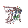
| ||||||||
|---|---|---|---|---|---|---|---|---|---|
| 1 |
| ||||||||
| Unit cell |
| ||||||||
| Components on special symmetry positions |
|
-Components
| #1: Protein | H3F3A Mass: 15257.816 Da / Num. of mol.: 1 / Mutation: S96A, Y99F, G102A, A111T, M120F Source method: isolated from a genetically manipulated source Source: (gene. exp.) Homo sapiens (human) / Gene: H3F3A, H3.3A, H3F3, PP781, H3F3B, H3.3B / Production host:  Escherichia coli (E. coli) / References: UniProt: P84243 Escherichia coli (E. coli) / References: UniProt: P84243 | ||
|---|---|---|---|
| #2: Protein | Mass: 11263.231 Da / Num. of mol.: 1 Source method: isolated from a genetically manipulated source Source: (gene. exp.) Homo sapiens (human)Gene: HIST1H4A, H4/A, H4FA, HIST1H4B, H4/I, H4FI, HIST1H4C, H4/G, H4FG, HIST1H4D, H4/B, H4FB, HIST1H4E, H4/J, H4FJ, HIST1H4F, H4/C, H4FC, HIST1H4H, H4/H, H4FH, HIST1H4I, H4/M, H4FM, HIST1H4J, H4/E, ...Gene: HIST1H4A, H4/A, H4FA, HIST1H4B, H4/I, H4FI, HIST1H4C, H4/G, H4FG, HIST1H4D, H4/B, H4FB, HIST1H4E, H4/J, H4FJ, HIST1H4F, H4/C, H4FC, HIST1H4H, H4/H, H4FH, HIST1H4I, H4/M, H4FM, HIST1H4J, H4/E, H4FE, HIST1H4K, H4/D, H4FD, HIST1H4L, H4/K, H4FK, HIST2H4A, H4/N, H4F2, H4FN, HIST2H4, HIST2H4B, H4/O, H4FO, HIST4H4 Production host: Escherichia coli (E. coli) / References: UniProt: P62805 | ||
| #3: Protein | Mass: 24725.328 Da / Num. of mol.: 1 / Fragment: UNP residues 178-389 Source method: isolated from a genetically manipulated source Source: (gene. exp.) Homo sapiens (human) / Gene: DAXX, BING2, DAP6 / Production host: Escherichia coli (E. coli) / References: UniProt: Q9UER7 | ||
| #4: Chemical | ChemComp-PO4 / Phosphate  Mass: 94.971 Da / Num. of mol.: 9 / Source method: obtained synthetically / Formula: PO4 Mass: 94.971 Da / Num. of mol.: 9 / Source method: obtained synthetically / Formula: PO4#5: Water | ChemComp-HOH / | Water Mass: 18.015 Da / Num. of mol.: 355 / Source method: isolated from a natural source / Formula: H2O Mass: 18.015 Da / Num. of mol.: 355 / Source method: isolated from a natural source / Formula: H2O |
-Experimental details
-Experiment
| Experiment | Method: X-RAY DIFFRACTION / Number of used crystals: 1 |
|---|
- Sample preparation
| Crystal | Density Matthews: 2.55 Å3/Da / Density % sol: 51.74 % |
|---|---|
| Crystal grow | Temperature: 277 K / Method: vapor diffusion, hanging drop / pH: 7 Details: 1.8 M Na/K-phosphate, pH 7.0, VAPOR DIFFUSION, HANGING DROP, temperature 277K |
-Data collection
| Diffraction source | Source: SYNCHROTRON / Site: NSLS  / Beamline: X29A / Wavelength: 0.979 Å / Beamline: X29A / Wavelength: 0.979 Å |
|---|---|
| Radiation | Protocol: SINGLE WAVELENGTH / Monochromatic (M) / Laue (L): M / Scattering type: x-ray |
| Radiation wavelength | Wavelength: 0.979 Å / Relative weight: 1 |
| Reflection | Resolution: 1.95→50 Å / Num. all: 39270 / Num. obs: 39270 / % possible obs: 99.9 % / Observed criterion σ(F): 2 / Observed criterion σ(I): 2 |
- Processing
| Software |
| |||||||||||||||||||||||||||||||||||||||||||||||||||||||||||||||||||||||||||||||||||||||||||||||||||||||||
|---|---|---|---|---|---|---|---|---|---|---|---|---|---|---|---|---|---|---|---|---|---|---|---|---|---|---|---|---|---|---|---|---|---|---|---|---|---|---|---|---|---|---|---|---|---|---|---|---|---|---|---|---|---|---|---|---|---|---|---|---|---|---|---|---|---|---|---|---|---|---|---|---|---|---|---|---|---|---|---|---|---|---|---|---|---|---|---|---|---|---|---|---|---|---|---|---|---|---|---|---|---|---|---|---|---|---|
| Refinement | Method to determine structure: MOLECULAR REPLACEMENT / Resolution: 1.95→48.058 Å / SU ML: 0.42 / σ(F): 1.36 / σ(I): 5.1 / Phase error: 20.42 / Stereochemistry target values: ML
| |||||||||||||||||||||||||||||||||||||||||||||||||||||||||||||||||||||||||||||||||||||||||||||||||||||||||
| Solvent computation | Shrinkage radii: 0.86 Å / VDW probe radii: 1.1 Å / Solvent model: FLAT BULK SOLVENT MODEL / Bsol: 44.673 Å2 / ksol: 0.364 e/Å3 | |||||||||||||||||||||||||||||||||||||||||||||||||||||||||||||||||||||||||||||||||||||||||||||||||||||||||
| Displacement parameters |
| |||||||||||||||||||||||||||||||||||||||||||||||||||||||||||||||||||||||||||||||||||||||||||||||||||||||||
| Refinement step | Cycle: LAST / Resolution: 1.95→48.058 Å
| |||||||||||||||||||||||||||||||||||||||||||||||||||||||||||||||||||||||||||||||||||||||||||||||||||||||||
| Refine LS restraints |
| |||||||||||||||||||||||||||||||||||||||||||||||||||||||||||||||||||||||||||||||||||||||||||||||||||||||||
| LS refinement shell |
|
