 Movie
Movie Controller

[English] 日本語
 Yorodumi
- PDB-4h3u: Crystal structure of hypothetical protein with ketosteroid isomer... -
+ Open data

- Basic information
| Entry | Database: PDB / ID: 4h3u | ||||||
|---|---|---|---|---|---|---|---|
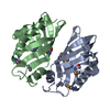
| Title | Crystal structure of hypothetical protein with ketosteroid isomerase-like protein fold from Catenulispora acidiphila DSM 44928 | ||||||
 Components Components | hypothetical protein | ||||||
 Keywords Keywords | Structural Genomics / Unknown Function / PSI-Biology / Midwest Center for Structural Genomics / MCSG | ||||||
| Function / homology |  Function and homology information Function and homology informationSnoaL-like domain / SnoaL-like domain / Nuclear Transport Factor 2; Chain: A, - #50 / NTF2-like domain superfamily / Nuclear Transport Factor 2; Chain: A, / Roll / Alpha Beta Similarity search - Domain/homology | ||||||
| Biological species |  Catenulispora acidiphila (bacteria) Catenulispora acidiphila (bacteria) | ||||||
| Method | X-RAY DIFFRACTION / SYNCHROTRON / SAD / Resolution: 1.15 Å | ||||||
 Authors Authors | Filippova, E.V. / Minasov, G. / Shuvalova, L. / Kiryukhina, O. / Jedrzejczak, R. / Joachimiak, A. / Anderson, W.F. / Midwest Center for Structural Genomics (MCSG) | ||||||
 Citation Citation | Journal: J.Struct.Funct.Genom. / Year: 2014 Title: Structural characterization of a hypothetical protein: a potential agent involved in trimethylamine metabolism in Catenulispora acidiphila. Authors: Filippova, E.V. / Luan, C.H. / Dunne, S.F. / Kiryukhina, O. / Minasov, G. / Shuvalova, L. / Anderson, W.F. | ||||||
| History |
|
- Structure visualization
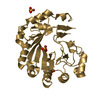
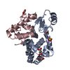
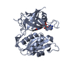
| Structure viewer | Molecule: MolmilJmol/JSmol |
|---|
- Downloads & links
-Download
| PDBx/mmCIF format | 4h3u.cif.gz | 134.1 KB | Display | PDBx/mmCIF format |
|---|---|---|---|---|
| PDB format | pdb4h3u.ent.gz | 111.2 KB | Display | PDB format |
| PDBx/mmJSON format | 4h3u.json.gz | Tree view | PDBx/mmJSON format | |
| Others |  Other downloads Other downloads |
-Validation report
| Arichive directory | https://data.pdbj.org/pub/pdb/validation_reports/h3/4h3uftp://data.pdbj.org/pub/pdb/validation_reports/h3/4h3u | HTTPS FTP |
|---|
-Related structure data


-Links
 PDBj
PDBj
- Assembly
| Deposited unit | 
| ||||||||
|---|---|---|---|---|---|---|---|---|---|
| 1 |
| ||||||||
| Unit cell |
|
-Components
| #1: Protein | Mass: 17246.256 Da / Num. of mol.: 2 Source method: isolated from a genetically manipulated source Source: (gene. exp.) Catenulispora acidiphila (bacteria) / Strain: DSM 44928 / Gene: Caci_0382 / Plasmid: pMCSG73 / Production host: Escherichia coli (E. coli) / Strain (production host): BL21(DE3)magic / References: UniProt: C7PVH9#2: Chemical | ChemComp-CD /   Mass: 112.411 Da / Num. of mol.: 7 / Source method: obtained synthetically / Formula: Cd Mass: 112.411 Da / Num. of mol.: 7 / Source method: obtained synthetically / Formula: Cd#3: Chemical | ChemComp-CL / Chloride  Mass: 35.453 Da / Num. of mol.: 7 / Source method: obtained synthetically / Formula: Cl Mass: 35.453 Da / Num. of mol.: 7 / Source method: obtained synthetically / Formula: Cl#4: Chemical | ChemComp-ACT / Acetate  Mass: 59.044 Da / Num. of mol.: 4 / Source method: obtained synthetically / Formula: C2H3O2 Mass: 59.044 Da / Num. of mol.: 4 / Source method: obtained synthetically / Formula: C2H3O2#5: Water | ChemComp-HOH / | Water Mass: 18.015 Da / Num. of mol.: 417 / Source method: isolated from a natural source / Formula: H2O Mass: 18.015 Da / Num. of mol.: 417 / Source method: isolated from a natural source / Formula: H2O |
|---|
-Experimental details
-Experiment
| Experiment | Method: X-RAY DIFFRACTION / Number of used crystals: 1 |
|---|
- Sample preparation
| Crystal | Density Matthews: 2.29 Å3/Da / Density % sol: 46.38 % |
|---|---|
| Crystal grow | Temperature: 293 K / Method: vapor diffusion, sitting drop / pH: 7.5 Details: 0.05 M Cadmium Sulfate, 0.1 M Hepes, 0.5 M Sodium acetate, pH 7.5, VAPOR DIFFUSION, SITTING DROP, temperature 293K |
-Data collection
| Diffraction | Mean temperature: 100 K |
|---|---|
| Diffraction source | Source: SYNCHROTRON / Site: APS  / Beamline: 21-ID-G / Wavelength: 0.97857 Å / Beamline: 21-ID-G / Wavelength: 0.97857 Å |
| Detector | Type: MARMOSAIC 300 mm CCD / Detector: CCD / Date: Aug 13, 2012 / Details: MIRROR |
| Radiation | Monochromator: SI-111 CHANNEL / Protocol: SINGLE WAVELENGTH / Monochromatic (M) / Laue (L): M / Scattering type: x-ray |
| Radiation wavelength | Wavelength: 0.97857 Å / Relative weight: 1 |
| Reflection | Resolution: 1.15→50 Å / Num. all: 212580 / Num. obs: 212580 / % possible obs: 97.4 % / Observed criterion σ(I): -3 / Redundancy: 5.8 % / Biso Wilson estimate: 14.6 Å2 / Rmerge(I) obs: 0.05 / Net I/σ(I): 34.7 |
| Reflection shell | Resolution: 1.15→1.17 Å / Redundancy: 5.6 % / Rmerge(I) obs: 0.5 / Mean I/σ(I) obs: 2.8 / Num. unique all: 10416 / % possible all: 95.7 |
- Processing
| Software |
| |||||||||||||||||||||||||||||||||||||||||||||||||||||||||||||||||||||||||||
|---|---|---|---|---|---|---|---|---|---|---|---|---|---|---|---|---|---|---|---|---|---|---|---|---|---|---|---|---|---|---|---|---|---|---|---|---|---|---|---|---|---|---|---|---|---|---|---|---|---|---|---|---|---|---|---|---|---|---|---|---|---|---|---|---|---|---|---|---|---|---|---|---|---|---|---|---|
| Refinement | Method to determine structure: SAD / Resolution: 1.15→29.94 Å / Cor.coef. Fo:Fc: 0.979 / Cor.coef. Fo:Fc free: 0.975 / SU B: 0.838 / SU ML: 0.018 / Isotropic thermal model: ANISOTROPIC / Cross valid method: THROUGHOUT / σ(F): 0 / ESU R: 0.027 / ESU R Free: 0.026 / Stereochemistry target values: MAXIMUM LIKELIHOOD / Details: HYDROGENS HAVE BEEN ADDED IN THE RIDING POSITIONS
| |||||||||||||||||||||||||||||||||||||||||||||||||||||||||||||||||||||||||||
| Solvent computation | Ion probe radii: 0.8 Å / Shrinkage radii: 0.8 Å / VDW probe radii: 1.2 Å / Solvent model: MASK | |||||||||||||||||||||||||||||||||||||||||||||||||||||||||||||||||||||||||||
| Displacement parameters | Biso mean: 14.577 Å2
| |||||||||||||||||||||||||||||||||||||||||||||||||||||||||||||||||||||||||||
| Refine analyze | Luzzati coordinate error obs: 0.1 Å | |||||||||||||||||||||||||||||||||||||||||||||||||||||||||||||||||||||||||||
| Refinement step | Cycle: LAST / Resolution: 1.15→29.94 Å
| |||||||||||||||||||||||||||||||||||||||||||||||||||||||||||||||||||||||||||
| Refine LS restraints |
| |||||||||||||||||||||||||||||||||||||||||||||||||||||||||||||||||||||||||||
| LS refinement shell | Resolution: 1.15→1.18 Å / Total num. of bins used: 20
| |||||||||||||||||||||||||||||||||||||||||||||||||||||||||||||||||||||||||||
| Refinement TLS params. | Method: refined / Refine-ID: X-RAY DIFFRACTION
| |||||||||||||||||||||||||||||||||||||||||||||||||||||||||||||||||||||||||||
| Refinement TLS group |
|
