Mass: 18.015 Da / Num. of mol.: 390 / Source method: isolated from a natural source / Formula: H2O
-
Details
Sequence details
AUTHORS STATE THAT, (1) IN CANDIDA ALBICANS RESIDUE 405 WOULD BE SERINE BECAUSE OF ITS CODON USAGE, ...AUTHORS STATE THAT, (1) IN CANDIDA ALBICANS RESIDUE 405 WOULD BE SERINE BECAUSE OF ITS CODON USAGE, BUT BECAUSE OF RECOMBINANT PROTEIN EXPRESSION IN E. COLI IT WAS TRANSLATED AS LEUCINE; (2) REGARDING RESIDUE 683, THERE MIGHT BE VARIATIONS FROM SOME OF THE PUBLISHED SEQUENCES DUE TO DIFFERENCES IN THE STRAIN OF YEAST SEQUENCED.
-
Experimental details
-
Experiment
Experiment
Method: X-RAY DIFFRACTION / Number of used crystals: 1
-
Sample preparation
Crystal
Density Matthews: 2.3 Å3/Da / Density % sol: 46.46 %
Crystal grow
Temperature: 277 K / Method: vapor diffusion, hanging drop / pH: 7.5 Details: 0.1 M Hepes, 14% PEG 4000, 75 mM NaCl, 5% ethylene glycol, pH 7.5, VAPOR DIFFUSION, HANGING DROP, temperature 277K
In the structure databanks used in Yorodumi, some data are registered as the other names, "COVID-19 virus" and "2019-nCoV". Here are the details of the virus and the list of structure data.
Jan 31, 2019. EMDB accession codes are about to change! (news from PDBe EMDB page)
EMDB accession codes are about to change! (news from PDBe EMDB page)
The allocation of 4 digits for EMDB accession codes will soon come to an end. Whilst these codes will remain in use, new EMDB accession codes will include an additional digit and will expand incrementally as the available range of codes is exhausted. The current 4-digit format prefixed with “EMD-” (i.e. EMD-XXXX) will advance to a 5-digit format (i.e. EMD-XXXXX), and so on. It is currently estimated that the 4-digit codes will be depleted around Spring 2019, at which point the 5-digit format will come into force.
The EM Navigator/Yorodumi systems omit the EMD- prefix.
Related info.:Q: What is EMD? / ID/Accession-code notation in Yorodumi/EM Navigator
Yorodumi is a browser for structure data from EMDB, PDB, SASBDB, etc.
This page is also the successor to EM Navigator detail page, and also detail information page/front-end page for Omokage search.
The word "yorodu" (or yorozu) is an old Japanese word meaning "ten thousand". "mi" (miru) is to see.
Related info.:EMDB / PDB / SASBDB / Comparison of 3 databanks / Yorodumi Search / Aug 31, 2016. New EM Navigator & Yorodumi / Yorodumi Papers / Jmol/JSmol / Function and homology information / Changes in new EM Navigator and Yorodumi
 Movie
Movie Controller

 Yorodumi
 Open data
 Basic information
 Components
 Keywords
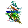
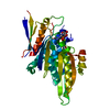
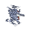
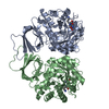
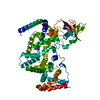
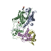
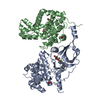
 MOTOR PROTEIN / Kinesin motor domain /
  Function and homology information

 Authors
 Citation
 Structure visualization
 Downloads & links
 Other downloads
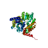


 PDBj
PDBj


 Assembly


 Mass: 24.305 Da / Num. of mol.: 1 / Source method: obtained synthetically / Formula: Mg
 Mass: 427.201 Da / Num. of mol.: 1 / Source method: obtained synthetically / Formula: C10H15N5O10P2 / Comment: ADP, energy-carrying molecule*YM
 Mass: 62.068 Da / Num. of mol.: 1 / Source method: obtained synthetically / Formula: C2H6O2
 Sample preparation
 / Beamline: 23-ID-B / Wavelength: 1.0332 Å
 Processing