 Movie
Movie Controller

[English] 日本語
 Yorodumi
- PDB-4gvf: Crystal structure of Salmonella typhimurium family 3 glycoside hy... -
+ Open data

- Basic information
| Entry | Database: PDB / ID: 4gvf | |||||||||
|---|---|---|---|---|---|---|---|---|---|---|
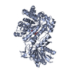
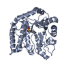
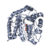
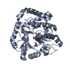
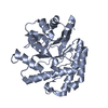
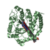
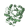
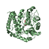
| Title | Crystal structure of Salmonella typhimurium family 3 glycoside hydrolase (NagZ) bound to GlcNAc | |||||||||
 Components Components | Beta-hexosaminidase Hexosaminidase Hexosaminidase | |||||||||
 Keywords Keywords | HYDROLASE / TIM-BARREL | |||||||||
| Function / homology |  Function and homology informationbeta-N-acetylhexosaminidase activity / beta-N-acetylhexosaminidase / peptidoglycan turnover / N-acetyl-beta-D-galactosaminidase activity / peptidoglycan biosynthetic process / cell wall organization / regulation of cell shape / carbohydrate metabolic process / cell cycle / cell division ...beta-N-acetylhexosaminidase activity / beta-N-acetylhexosaminidase / peptidoglycan turnover / N-acetyl-beta-D-galactosaminidase activity / peptidoglycan biosynthetic process / cell wall organization / regulation of cell shape / carbohydrate metabolic process / cell cycle / cell division / response to antibiotic / cytoplasm Function and homology informationbeta-N-acetylhexosaminidase activity / beta-N-acetylhexosaminidase / peptidoglycan turnover / N-acetyl-beta-D-galactosaminidase activity / peptidoglycan biosynthetic process / cell wall organization / regulation of cell shape / carbohydrate metabolic process / cell cycle / cell division ...beta-N-acetylhexosaminidase activity / beta-N-acetylhexosaminidase / peptidoglycan turnover / N-acetyl-beta-D-galactosaminidase activity / peptidoglycan biosynthetic process / cell wall organization / regulation of cell shape / carbohydrate metabolic process / cell cycle / cell division / response to antibiotic / cytoplasmSimilarity search - Function | |||||||||
| Biological species |  Salmonella enterica subsp. enterica serovar Typhimurium (bacteria) Salmonella enterica subsp. enterica serovar Typhimurium (bacteria) | |||||||||
| Method | X-RAY DIFFRACTION / SYNCHROTRON / MOLECULAR REPLACEMENT / Resolution: 1.35 Å | |||||||||
 Authors Authors | Bacik, J.P. / Mark, B.L. | |||||||||
 Citation Citation | Journal: Chem.Biol. / Year: 2012 Title: Active Site Plasticity within the Glycoside Hydrolase NagZ Underlies a Dynamic Mechanism of Substrate Distortion. Authors: Bacik, J.P. / Whitworth, G.E. / Stubbs, K.A. / Vocadlo, D.J. / Mark, B.L. | |||||||||
| History |
|
- Structure visualization
| Structure viewer | Molecule: MolmilJmol/JSmol |
|---|
- Downloads & links
-Download
| PDBx/mmCIF format | 4gvf.cif.gz | 163.5 KB | Display | PDBx/mmCIF format |
|---|---|---|---|---|
| PDB format | pdb4gvf.ent.gz | 128 KB | Display | PDB format |
| PDBx/mmJSON format | 4gvf.json.gz | Tree view | PDBx/mmJSON format | |
| Others |  Other downloads Other downloads |
-Validation report
| Arichive directory | https://data.pdbj.org/pub/pdb/validation_reports/gv/4gvfftp://data.pdbj.org/pub/pdb/validation_reports/gv/4gvf | HTTPS FTP |
|---|
-Related structure data


-Links
 PDBj
PDBj


- Assembly
| Deposited unit | 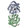
| ||||||||
|---|---|---|---|---|---|---|---|---|---|
| 1 | 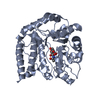
| ||||||||
| 2 | 
| ||||||||
| Unit cell |
|
-Components
| #1: Protein | Hexosaminidase / Beta-N-acetylhexosaminidase / N-acetyl-beta-glucosaminidase Mass: 38723.934 Da / Num. of mol.: 2 Source method: isolated from a genetically manipulated source Source: (gene. exp.) Salmonella enterica subsp. enterica serovar Typhimurium (bacteria)Strain: LT2 / SGSC1412 / ATCC 700720 / Gene: nagZ, STM1209 / Production host: Escherichia coli (E. coli) / References: UniProt: Q8ZQ06, beta-N-acetylhexosaminidase#2: Sugar | N-Acetylglucosamine  Type: D-saccharide, alpha linking / Mass: 221.208 Da / Num. of mol.: 2 Type: D-saccharide, alpha linking / Mass: 221.208 Da / Num. of mol.: 2Source method: isolated from a genetically manipulated source Formula: C8H15NO6 #3: Sugar | N-Acetylglucosamine  Type: D-saccharide, beta linking / Mass: 221.208 Da / Num. of mol.: 2 Type: D-saccharide, beta linking / Mass: 221.208 Da / Num. of mol.: 2Source method: isolated from a genetically manipulated source Formula: C8H15NO6 #4: Chemical | ChemComp-MES / | MES (buffer)  Mass: 195.237 Da / Num. of mol.: 1 / Source method: obtained synthetically / Formula: C6H13NO4S / Comment: pH buffer*YM Mass: 195.237 Da / Num. of mol.: 1 / Source method: obtained synthetically / Formula: C6H13NO4S / Comment: pH buffer*YM#5: Water | ChemComp-HOH / | Water Mass: 18.015 Da / Num. of mol.: 988 / Source method: isolated from a natural source / Formula: H2O Mass: 18.015 Da / Num. of mol.: 988 / Source method: isolated from a natural source / Formula: H2O |
|---|
-Experimental details
-Experiment
| Experiment | Method: X-RAY DIFFRACTION / Number of used crystals: 1 |
|---|
- Sample preparation
| Crystal | Density Matthews: 1.99 Å3/Da / Density % sol: 38.05 % |
|---|---|
| Crystal grow | Temperature: 296 K / pH: 6.5 Details: 0.1M MES, 25% PEG 1000, pH 6.5, VAPOR DIFFUSION, HANGING DROP, temperature 296K |
-Data collection
| Diffraction | Mean temperature: 100 K |
|---|---|
| Diffraction source | Source: SYNCHROTRON / Site: CLSI  / Beamline: 08ID-1 / Wavelength: 0.9795 / Beamline: 08ID-1 / Wavelength: 0.9795 |
| Detector | Type: RAYONIX MX-300 / Detector: CCD / Date: Mar 19, 2010 |
| Radiation | Protocol: SINGLE WAVELENGTH / Monochromatic (M) / Laue (L): M / Scattering type: x-ray |
| Radiation wavelength | Wavelength: 0.9795 Å / Relative weight: 1 |
| Reflection | Resolution: 1.35→48.89 Å / Num. obs: 131665 / % possible obs: 99.1 % / Observed criterion σ(I): 3 / Redundancy: 3.6 % / Biso Wilson estimate: 9.14 Å2 / Rmerge(I) obs: 0.06 / Rsym value: 0.07 / Net I/σ(I): 13.8 |
| Reflection shell | Resolution: 1.35→1.42 Å / Redundancy: 2.8 % / Rmerge(I) obs: 0.234 / Mean I/σ(I) obs: 4.7 / Rsym value: 0.29 / % possible all: 93.7 |
- Processing
| Software |
| |||||||||||||||||||||||||||||||||||||||||||||||||||||||||||||||||||||||||||||
|---|---|---|---|---|---|---|---|---|---|---|---|---|---|---|---|---|---|---|---|---|---|---|---|---|---|---|---|---|---|---|---|---|---|---|---|---|---|---|---|---|---|---|---|---|---|---|---|---|---|---|---|---|---|---|---|---|---|---|---|---|---|---|---|---|---|---|---|---|---|---|---|---|---|---|---|---|---|---|
| Refinement | Method to determine structure: MOLECULAR REPLACEMENT / Resolution: 1.35→48.89 Å / SU ML: 0.16 / σ(F): 0 / Phase error: 17.88 / Stereochemistry target values: ML
| |||||||||||||||||||||||||||||||||||||||||||||||||||||||||||||||||||||||||||||
| Solvent computation | Shrinkage radii: 0.72 Å / VDW probe radii: 1 Å / Solvent model: FLAT BULK SOLVENT MODEL / Bsol: 43.63 Å2 / ksol: 0.35 e/Å3 | |||||||||||||||||||||||||||||||||||||||||||||||||||||||||||||||||||||||||||||
| Displacement parameters |
| |||||||||||||||||||||||||||||||||||||||||||||||||||||||||||||||||||||||||||||
| Refinement step | Cycle: LAST / Resolution: 1.35→48.89 Å
| |||||||||||||||||||||||||||||||||||||||||||||||||||||||||||||||||||||||||||||
| Refine LS restraints |
| |||||||||||||||||||||||||||||||||||||||||||||||||||||||||||||||||||||||||||||
| LS refinement shell |
|
