 Movie
Movie Controller

[English] 日本語
 Yorodumi
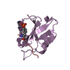
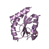
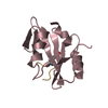
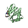
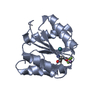
- PDB-4gof: Crystal structure of the SGTA homodimerization domain with covale... -
+ Open data

- Basic information
| Entry | Database: PDB / ID: 4gof | ||||||
|---|---|---|---|---|---|---|---|
| Title | Crystal structure of the SGTA homodimerization domain with covalent modifications to both C38 | ||||||
 Components Components | Small glutamine-rich tetratricopeptide repeat-containing protein alpha | ||||||
 Keywords Keywords |  PROTEIN BINDING / Four-helix bundle / Protein-protein interaction / Ubl4A ubiquitin-like domain PROTEIN BINDING / Four-helix bundle / Protein-protein interaction / Ubl4A ubiquitin-like domain | ||||||
| Function / homology |  Function and homology information Function and homology informationTRC complex / negative regulation of ERAD pathway / positive regulation of ERAD pathway / tail-anchored membrane protein insertion into ER membrane / post-translational protein targeting to endoplasmic reticulum membrane / positive regulation of ubiquitin-dependent protein catabolic process / BAT3 complex binding / Insertion of tail-anchored proteins into the endoplasmic reticulum membrane / ERAD pathway / negative regulation of ubiquitin-dependent protein catabolic process ...TRC complex / negative regulation of ERAD pathway / positive regulation of ERAD pathway / tail-anchored membrane protein insertion into ER membrane / post-translational protein targeting to endoplasmic reticulum membrane / positive regulation of ubiquitin-dependent protein catabolic process / BAT3 complex binding / Insertion of tail-anchored proteins into the endoplasmic reticulum membrane / ERAD pathway / negative regulation of ubiquitin-dependent protein catabolic process / : / molecular adaptor activity / membrane / identical protein binding / nucleus / cytosol / cytoplasmSimilarity search - Function | ||||||
| Biological species |  Homo sapiens (human) Homo sapiens (human) | ||||||
| Method | X-RAY DIFFRACTION / SYNCHROTRON / MOLECULAR REPLACEMENT / Resolution: 1.35 Å | ||||||
 Authors Authors | Chartron, J.W. / VanderVelde, D.G. / Clemons Jr., W.M. | ||||||
 Citation Citation | Journal: Cell Rep / Year: 2012 Title: Structures of the Sgt2/SGTA Dimerization Domain with the Get5/UBL4A UBL Domain Reveal an Interaction that Forms a Conserved Dynamic Interface. Authors: Chartron, J.W. / Vandervelde, D.G. / Clemons, W.M. | ||||||
| History |
|
- Structure visualization
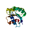
| Structure viewer | Molecule: MolmilJmol/JSmol |
|---|
- Downloads & links
-Download
| PDBx/mmCIF format | 4gof.cif.gz | 29.8 KB | Display | PDBx/mmCIF format |
|---|---|---|---|---|
| PDB format | pdb4gof.ent.gz | 23 KB | Display | PDB format |
| PDBx/mmJSON format | 4gof.json.gz | Tree view | PDBx/mmJSON format | |
| Others |  Other downloads Other downloads |
-Validation report
| Arichive directory | https://data.pdbj.org/pub/pdb/validation_reports/go/4gofftp://data.pdbj.org/pub/pdb/validation_reports/go/4gof | HTTPS FTP |
|---|
-Related structure data
| Related structure data |  2lxaC 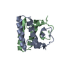 2lxbC  2lxcC  4gocC  4godC  4goeC C: citing same article ( |
|---|---|
| Similar structure data |


-Links
 PDBj
PDBj


- Assembly
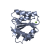
| Deposited unit | 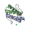
| |||||||||||||||
|---|---|---|---|---|---|---|---|---|---|---|---|---|---|---|---|---|
| 1 |
| |||||||||||||||
| Unit cell |
| |||||||||||||||
| Components on special symmetry positions |
|
-Components
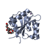
| #1: Protein | Mass: 5738.505 Da / Num. of mol.: 2 Source method: isolated from a genetically manipulated source Source: (gene. exp.) Homo sapiens (human) / Gene: SGT, SGT1, SGTA / Plasmid: pET33b / Production host:  Escherichia coli (E. coli) / Strain (production host): NiCo21(DE3) / References: UniProt: O43765 Escherichia coli (E. coli) / Strain (production host): NiCo21(DE3) / References: UniProt: O43765#2: Chemical | 2-Mercaptoethanol  Mass: 78.133 Da / Num. of mol.: 2 / Source method: obtained synthetically / Formula: C2H6OS Mass: 78.133 Da / Num. of mol.: 2 / Source method: obtained synthetically / Formula: C2H6OS#3: Chemical | ChemComp-CL / | Chloride  Mass: 35.453 Da / Num. of mol.: 1 / Source method: obtained synthetically / Formula: Cl Mass: 35.453 Da / Num. of mol.: 1 / Source method: obtained synthetically / Formula: Cl#4: Water | ChemComp-HOH / | Water Mass: 18.015 Da / Num. of mol.: 81 / Source method: isolated from a natural source / Formula: H2O Mass: 18.015 Da / Num. of mol.: 81 / Source method: isolated from a natural source / Formula: H2O |
|---|
-Experimental details
-Experiment
| Experiment | Method: X-RAY DIFFRACTION / Number of used crystals: 1 |
|---|
- Sample preparation
| Crystal | Density Matthews: 1.78 Å3/Da / Density % sol: 31.09 % |
|---|---|
| Crystal grow | Temperature: 277 K / Method: vapor diffusion, sitting drop / pH: 5 Details: 10% 2-propanol, 0.1 M sodium citrate, 26% PEG 400, 10 mM 2-mercaptoethanol, pH 5.0, VAPOR DIFFUSION, SITTING DROP, temperature 277K |
-Data collection
| Diffraction | Mean temperature: 100 K |
|---|---|
| Diffraction source | Source: SYNCHROTRON / Site: SSRL  / Beamline: BL12-2 / Wavelength: 1 Å / Beamline: BL12-2 / Wavelength: 1 Å |
| Detector | Type: PSI PILATUS 6M / Detector: PIXEL / Date: Feb 9, 2012 |
| Radiation | Monochromator: Liquid nitrogen-cooled double crystal / Protocol: SINGLE WAVELENGTH / Monochromatic (M) / Laue (L): M / Scattering type: x-ray |
| Radiation wavelength | Wavelength: 1 Å / Relative weight: 1 |
| Reflection | Resolution: 1.35→19.39 Å / Num. obs: 18421 / % possible obs: 98.9 % / Redundancy: 3.9 % / Rmerge(I) obs: 0.058 / Net I/σ(I): 10.1 |
| Reflection shell | Resolution: 1.35→1.37 Å / Redundancy: 3.6 % / Rmerge(I) obs: 0.692 / Mean I/σ(I) obs: 1.7 / Num. unique all: 876 / % possible all: 99.6 |
- Processing
| Software |
| |||||||||||||||||||||||||||||||||||||||||||||||||||||||||||||||
|---|---|---|---|---|---|---|---|---|---|---|---|---|---|---|---|---|---|---|---|---|---|---|---|---|---|---|---|---|---|---|---|---|---|---|---|---|---|---|---|---|---|---|---|---|---|---|---|---|---|---|---|---|---|---|---|---|---|---|---|---|---|---|---|---|
| Refinement | Method to determine structure: MOLECULAR REPLACEMENT / Resolution: 1.35→19.39 Å / SU ML: 0.13 / σ(F): 1.77 / Phase error: 18.8 / Stereochemistry target values: ML
| |||||||||||||||||||||||||||||||||||||||||||||||||||||||||||||||
| Solvent computation | Shrinkage radii: 0.9 Å / VDW probe radii: 1.11 Å / Solvent model: FLAT BULK SOLVENT MODEL | |||||||||||||||||||||||||||||||||||||||||||||||||||||||||||||||
| Refinement step | Cycle: LAST / Resolution: 1.35→19.39 Å
| |||||||||||||||||||||||||||||||||||||||||||||||||||||||||||||||
| Refine LS restraints |
| |||||||||||||||||||||||||||||||||||||||||||||||||||||||||||||||
| LS refinement shell |
|
