 Movie
Movie Controller

[English] 日本語
 Yorodumi
- PDB-4ghe: Structure of Y257F variant of Homoprotocatechuate 2,3-Dioxygenase... -
+ Open data

- Basic information
| Entry | Database: PDB / ID: 4ghe | ||||||
|---|---|---|---|---|---|---|---|
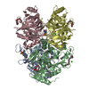
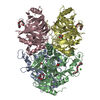
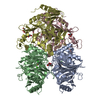
| Title | Structure of Y257F variant of Homoprotocatechuate 2,3-Dioxygenase from B.fuscum in complex with 4-nitrocatechol at 1.60 Ang resolution | ||||||
 Components Components | Homoprotocatechuate 2,3-dioxygenase | ||||||
 Keywords Keywords |  OXIDOREDUCTASE / Dioxygenase / oxygen activation / Fe(II) / 2-His-1-carboxylate facial triad / homoprotocatechuate / 4-nitrocatechol / oxy complex OXIDOREDUCTASE / Dioxygenase / oxygen activation / Fe(II) / 2-His-1-carboxylate facial triad / homoprotocatechuate / 4-nitrocatechol / oxy complex | ||||||
| Function / homology |  Function and homology information Function and homology information | ||||||
| Biological species |  Brevibacterium fuscum (bacteria) Brevibacterium fuscum (bacteria) | ||||||
| Method | X-RAY DIFFRACTION / SYNCHROTRON / MOLECULAR REPLACEMENT / Resolution: 1.6 Å | ||||||
 Authors Authors | Kovaleva, E.G. / Lipscomb, J.D. | ||||||
 Citation Citation | Journal: Biochemistry / Year: 2012 Title: Structural basis for the role of tyrosine 257 of homoprotocatechuate 2,3-dioxygenase in substrate and oxygen activation. Authors: Kovaleva, E.G. / Lipscomb, J.D. | ||||||
| History |
|
- Structure visualization
| Structure viewer | Molecule: MolmilJmol/JSmol |
|---|
- Downloads & links
-Download
| PDBx/mmCIF format | 4ghe.cif.gz | 611.8 KB | Display | PDBx/mmCIF format |
|---|---|---|---|---|
| PDB format | pdb4ghe.ent.gz | 505.2 KB | Display | PDB format |
| PDBx/mmJSON format | 4ghe.json.gz | Tree view | PDBx/mmJSON format | |
| Others |  Other downloads Other downloads |
-Validation report
| Arichive directory | https://data.pdbj.org/pub/pdb/validation_reports/gh/4gheftp://data.pdbj.org/pub/pdb/validation_reports/gh/4ghe | HTTPS FTP |
|---|
-Related structure data
| Related structure data |  4ghcC  4ghdC  4ghfC  4ghgC  4ghhC  3ojtS C: citing same article ( S: Starting model for refinement |
|---|---|
| Similar structure data |


-Links
 PDBj
PDBj


- Assembly
| Deposited unit | 
| |||||||||
|---|---|---|---|---|---|---|---|---|---|---|
| 1 |
| |||||||||
| Unit cell |
| |||||||||
| Components on special symmetry positions |
|
-Components
-Protein , 1 types, 4 molecules ABCD
| #1: Protein | Mass: 41739.320 Da / Num. of mol.: 4 / Mutation: Y257F Source method: isolated from a genetically manipulated source Source: (gene. exp.) Brevibacterium fuscum (bacteria) / Strain: ATCC 15993 / Gene: hpcd / Plasmid: pYZW204 / Production host: Escherichia coli (E. coli) / Strain (production host): JM109References: UniProt: Q45135, 3,4-dihydroxyphenylacetate 2,3-dioxygenase |
|---|
-Non-polymers , 6 types, 1624 molecules 


| #2: Chemical | ChemComp-FE2 /  Mass: 55.845 Da / Num. of mol.: 4 / Source method: obtained synthetically / Formula: Fe Mass: 55.845 Da / Num. of mol.: 4 / Source method: obtained synthetically / Formula: Fe#3: Chemical | ChemComp-P6G / Polyethylene glycol Mass: 282.331 Da / Num. of mol.: 7 / Source method: obtained synthetically / Formula: C12H26O7 / Comment: precipitant*YM Mass: 282.331 Da / Num. of mol.: 7 / Source method: obtained synthetically / Formula: C12H26O7 / Comment: precipitant*YM#4: Chemical | ChemComp-CL / Chloride Mass: 35.453 Da / Num. of mol.: 4 / Source method: obtained synthetically / Formula: Cl Mass: 35.453 Da / Num. of mol.: 4 / Source method: obtained synthetically / Formula: Cl#5: Chemical | ChemComp-CA / |  Mass: 40.078 Da / Num. of mol.: 1 / Source method: obtained synthetically / Formula: Ca Mass: 40.078 Da / Num. of mol.: 1 / Source method: obtained synthetically / Formula: Ca#6: Chemical | ChemComp-4NC / |  Mass: 155.108 Da / Num. of mol.: 1 / Source method: obtained synthetically / Formula: C6H5NO4 Mass: 155.108 Da / Num. of mol.: 1 / Source method: obtained synthetically / Formula: C6H5NO4#7: Water | ChemComp-HOH / | WaterMass: 18.015 Da / Num. of mol.: 1607 / Source method: isolated from a natural source / Formula: H2O |
|---|
-Experimental details
-Experiment
| Experiment | Method: X-RAY DIFFRACTION / Number of used crystals: 1 |
|---|
- Sample preparation
| Crystal | Density Matthews: 2.39 Å3/Da / Density % sol: 48.51 % |
|---|---|
| Crystal grow | Temperature: 293 K / Method: vapor diffusion, hanging drop / pH: 6.5 Details: 13% PEG6000, 0.1M calcium chloride, 0.1M Tris-HCl. Cryoprotectant 25% PEG400. Ligand soaking: 2mM 4-nitrocatechol for 40min in anaerobic glovebox atmosphere prior to cryo-cooling in liquid ...Details: 13% PEG6000, 0.1M calcium chloride, 0.1M Tris-HCl. Cryoprotectant 25% PEG400. Ligand soaking: 2mM 4-nitrocatechol for 40min in anaerobic glovebox atmosphere prior to cryo-cooling in liquid nitrogen. , pH 6.5, VAPOR DIFFUSION, HANGING DROP, temperature 293K |
-Data collection
| Diffraction | Mean temperature: 100 K |
|---|---|
| Diffraction source | Source: SYNCHROTRON / Site: SOLEIL  / Beamline: PROXIMA 1 / Wavelength: 0.9801 Å / Beamline: PROXIMA 1 / Wavelength: 0.9801 Å |
| Detector | Type: ADSC QUANTUM 315r / Detector: CCD / Date: Dec 11, 2010 |
| Radiation | Monochromator: Channel cut Si(111) / Protocol: SINGLE WAVELENGTH / Monochromatic (M) / Laue (L): M / Scattering type: x-ray |
| Radiation wavelength | Wavelength: 0.9801 Å / Relative weight: 1 |
| Reflection | Resolution: 1.6→42.3 Å / Num. all: 207338 / Num. obs: 207338 / % possible obs: 98.8 % / Observed criterion σ(F): 0 / Observed criterion σ(I): -3 / Redundancy: 3.3 % / Rmerge(I) obs: 0.041 / Net I/σ(I): 12.8 |
| Reflection shell | Resolution: 1.6→1.69 Å / Redundancy: 3.3 % / Rmerge(I) obs: 0.407 / Mean I/σ(I) obs: 2.1 / Num. unique all: 30148 / % possible all: 99.1 |
- Processing
| Software |
| |||||||||||||||||||||||||||||||||||||||||||||||||||||||||||||||||||||||||||||||||||||||||||||||||||||||||||||||||||||||||||||
|---|---|---|---|---|---|---|---|---|---|---|---|---|---|---|---|---|---|---|---|---|---|---|---|---|---|---|---|---|---|---|---|---|---|---|---|---|---|---|---|---|---|---|---|---|---|---|---|---|---|---|---|---|---|---|---|---|---|---|---|---|---|---|---|---|---|---|---|---|---|---|---|---|---|---|---|---|---|---|---|---|---|---|---|---|---|---|---|---|---|---|---|---|---|---|---|---|---|---|---|---|---|---|---|---|---|---|---|---|---|---|---|---|---|---|---|---|---|---|---|---|---|---|---|---|---|---|
| Refinement | Method to determine structure: MOLECULAR REPLACEMENT Starting model: 3OJT Resolution: 1.6→42.3 Å / Cor.coef. Fo:Fc: 0.972 / Cor.coef. Fo:Fc free: 0.96 / SU B: 3.246 / SU ML: 0.058 / Cross valid method: THROUGHOUT / ESU R: 0.078 / ESU R Free: 0.08 / Stereochemistry target values: MAXIMUM LIKELIHOOD / Details: HYDROGENS HAVE BEEN ADDED IN THE RIDING POSITIONS
| |||||||||||||||||||||||||||||||||||||||||||||||||||||||||||||||||||||||||||||||||||||||||||||||||||||||||||||||||||||||||||||
| Solvent computation | Ion probe radii: 0.8 Å / Shrinkage radii: 0.8 Å / VDW probe radii: 1.2 Å / Solvent model: BABINET MODEL WITH MASK | |||||||||||||||||||||||||||||||||||||||||||||||||||||||||||||||||||||||||||||||||||||||||||||||||||||||||||||||||||||||||||||
| Displacement parameters | Biso mean: 22.436 Å2
| |||||||||||||||||||||||||||||||||||||||||||||||||||||||||||||||||||||||||||||||||||||||||||||||||||||||||||||||||||||||||||||
| Refinement step | Cycle: LAST / Resolution: 1.6→42.3 Å
| |||||||||||||||||||||||||||||||||||||||||||||||||||||||||||||||||||||||||||||||||||||||||||||||||||||||||||||||||||||||||||||
| Refine LS restraints |
| |||||||||||||||||||||||||||||||||||||||||||||||||||||||||||||||||||||||||||||||||||||||||||||||||||||||||||||||||||||||||||||
| LS refinement shell | Resolution: 1.6→1.642 Å / Total num. of bins used: 20
| |||||||||||||||||||||||||||||||||||||||||||||||||||||||||||||||||||||||||||||||||||||||||||||||||||||||||||||||||||||||||||||
| Refinement TLS params. | Method: refined / Refine-ID: X-RAY DIFFRACTION
| |||||||||||||||||||||||||||||||||||||||||||||||||||||||||||||||||||||||||||||||||||||||||||||||||||||||||||||||||||||||||||||
| Refinement TLS group |
|
