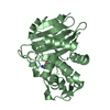
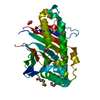
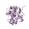
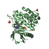
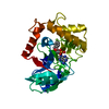
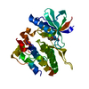
[English] 日本語
Yorodumi
- PDB-4gfu: PTPN18 in complex with HER2-pY1248 phosphor-peptides -

+
Open data


ID or keywords:

Loading...

-
Basic information

Entry
Database: PDB / ID: 4gfu
TitlePTPN18 in complex with HER2-pY1248 phosphor-peptides
Components
  • HER2-pY1248 phosphor-peptide
  • Tyrosine-protein phosphatase non-receptor type 18
KeywordsHYDROLASE/Peptide / phosphatase / tyrosine phosphorylation / hydrolase / HYDROLASE-Peptide complex
Function / homology
Function and homology information


negative regulation of ERBB signaling pathway / negative regulation of immature T cell proliferation in thymus / ERBB3:ERBB2 complex / ERBB2-ERBB4 signaling pathway / GRB7 events in ERBB2 signaling / immature T cell proliferation in thymus / RNA polymerase I core binding / regulation of microtubule-based process / blastocyst formation / ErbB-3 class receptor binding ...negative regulation of ERBB signaling pathway / negative regulation of immature T cell proliferation in thymus / ERBB3:ERBB2 complex / ERBB2-ERBB4 signaling pathway / GRB7 events in ERBB2 signaling / immature T cell proliferation in thymus / RNA polymerase I core binding / regulation of microtubule-based process / blastocyst formation / ErbB-3 class receptor binding / semaphorin receptor complex / Sema4D induced cell migration and growth-cone collapse / motor neuron axon guidance / Interleukin-37 signaling / neurotransmitter receptor localization to postsynaptic specialization membrane / PLCG1 events in ERBB2 signaling / ERBB2-EGFR signaling pathway / neuromuscular junction development / positive regulation of Rho protein signal transduction / ERBB2 Activates PTK6 Signaling / Drug-mediated inhibition of ERBB2 signaling / Resistance of ERBB2 KD mutants to trastuzumab / Resistance of ERBB2 KD mutants to sapitinib / Resistance of ERBB2 KD mutants to tesevatinib / Resistance of ERBB2 KD mutants to neratinib / Resistance of ERBB2 KD mutants to osimertinib / Resistance of ERBB2 KD mutants to afatinib / Resistance of ERBB2 KD mutants to AEE788 / Resistance of ERBB2 KD mutants to lapatinib / Drug resistance in ERBB2 TMD/JMD mutants / enzyme-linked receptor protein signaling pathway / positive regulation of transcription by RNA polymerase I / ERBB2-ERBB3 signaling pathway / oligodendrocyte differentiation / ERBB2 Regulates Cell Motility / semaphorin-plexin signaling pathway / PI3K events in ERBB2 signaling / non-membrane spanning protein tyrosine phosphatase activity / positive regulation of cell adhesion / positive regulation of protein targeting to membrane / regulation of angiogenesis / coreceptor activity / Schwann cell development / Signaling by ERBB2 / cellular response to epidermal growth factor stimulus / myelination / Downregulation of ERBB2:ERBB3 signaling / GRB2 events in ERBB2 signaling / TFAP2 (AP-2) family regulates transcription of growth factors and their receptors / transmembrane receptor protein tyrosine kinase activity / SHC1 events in ERBB2 signaling / Constitutive Signaling by Overexpressed ERBB2 / neurogenesis / protein dephosphorylation / protein-tyrosine-phosphatase / basal plasma membrane / regulation of ERK1 and ERK2 cascade / phosphatidylinositol 3-kinase/protein kinase B signal transduction / protein tyrosine phosphatase activity / positive regulation of translation / positive regulation of epithelial cell proliferation / cell surface receptor protein tyrosine kinase signaling pathway / Signaling by ERBB2 TMD/JMD mutants / positive regulation of MAP kinase activity / wound healing / neuromuscular junction / Signaling by ERBB2 ECD mutants / neuron differentiation / Signaling by ERBB2 KD Mutants / receptor protein-tyrosine kinase / receptor tyrosine kinase binding / cellular response to growth factor stimulus / Downregulation of ERBB2 signaling / ruffle membrane / peptidyl-tyrosine phosphorylation / Constitutive Signaling by Aberrant PI3K in Cancer / transmembrane signaling receptor activity / PIP3 activates AKT signaling / myelin sheath / presynaptic membrane / heart development / PI5P, PP2A and IER3 Regulate PI3K/AKT Signaling / RAF/MAP kinase cascade / positive regulation of cell growth / basolateral plasma membrane / protein tyrosine kinase activity / positive regulation of MAPK cascade / early endosome / cell surface receptor signaling pathway / receptor complex / endosome membrane / intracellular signal transduction / apical plasma membrane / positive regulation of protein phosphorylation / protein heterodimerization activity / protein phosphorylation / signaling receptor binding / positive regulation of cell population proliferation / negative regulation of apoptotic process / perinuclear region of cytoplasm
Similarity search - Function
: / : / : / Epidermal growth factor receptor transmembrane-juxtamembrane segment / Tyrosine protein kinase, EGF/ERB/XmrK receptor / Growth factor receptor domain 4 / Growth factor receptor domain IV / Receptor L-domain / Furin-like cysteine-rich domain / Receptor L-domain superfamily ...: / : / : / Epidermal growth factor receptor transmembrane-juxtamembrane segment / Tyrosine protein kinase, EGF/ERB/XmrK receptor / Growth factor receptor domain 4 / Growth factor receptor domain IV / Receptor L-domain / Furin-like cysteine-rich domain / Receptor L-domain superfamily / Furin-like cysteine rich region / Receptor L domain / Protein tyrosine phosphatase superfamily / Furin-like repeat / Furin-like repeats / Protein-Tyrosine Phosphatase; Chain A / Protein tyrosine phosphatase, catalytic domain / PTP type protein phosphatase domain profile. / Protein-tyrosine phosphatase / Tyrosine-specific protein phosphatase, PTPase domain / Protein-tyrosine phosphatase, catalytic / Protein tyrosine phosphatase, catalytic domain motif / Tyrosine specific protein phosphatases active site. / Protein-tyrosine phosphatase, active site / Tyrosine-specific protein phosphatases domain / Tyrosine specific protein phosphatases domain profile. / Protein-tyrosine phosphatase-like / Growth factor receptor cysteine-rich domain superfamily / Tyrosine-protein kinase, catalytic domain / Tyrosine kinase, catalytic domain / Tyrosine protein kinases specific active-site signature. / Tyrosine-protein kinase, active site / Protein tyrosine and serine/threonine kinase / Serine-threonine/tyrosine-protein kinase, catalytic domain / Protein kinase, ATP binding site / Protein kinases ATP-binding region signature. / Protein kinase domain profile. / Protein kinase domain / Protein kinase-like domain superfamily / Alpha-Beta Complex / Alpha Beta
Similarity search - Domain/homology
Receptor tyrosine-protein kinase erbB-2 / Tyrosine-protein phosphatase non-receptor type 18
Similarity search - Component
Biological speciesHomo sapiens (human)
MethodX-RAY DIFFRACTION / SYNCHROTRON / MOLECULAR REPLACEMENT / Resolution: 2 Å
AuthorsWang, H.M. / Yang, F. / Du, Y.J. / Yang, D.X. / Zhang, Y. / Yu, X. / Sun, J.P.
CitationJournal: To be Published
Title: PTPN18-HER2 peptides
Authors: Wang, H.M. / Yang, F. / Du, Y.J. / Yang, D.X. / Zhang, Y. / Yu, X. / Sun, J.P.
History
DepositionAug 4, 2012Deposition site: RCSB / Processing site: RCSB
Revision 1.0Aug 7, 2013Provider: repository / Type: Initial release
Revision 1.1Sep 13, 2023Group: Data collection / Database references ...Data collection / Database references / Derived calculations / Refinement description
Category: chem_comp_atom / chem_comp_bond ...chem_comp_atom / chem_comp_bond / database_2 / pdbx_initial_refinement_model / struct_conn / struct_ref_seq_dif
Item: _database_2.pdbx_DOI / _database_2.pdbx_database_accession ..._database_2.pdbx_DOI / _database_2.pdbx_database_accession / _struct_conn.pdbx_leaving_atom_flag / _struct_ref_seq_dif.details
Revision 1.2Dec 6, 2023Group: Data collection / Category: chem_comp_atom / chem_comp_bond / Item: _chem_comp_atom.atom_id / _chem_comp_bond.atom_id_2

-
Structure visualization

Structure viewerMolecule:
MolmilJmol/JSmol

Downloads & links

-
Assembly

Deposited unit
A: Tyrosine-protein phosphatase non-receptor type 18
F: HER2-pY1248 phosphor-peptide


Theoretical massNumber of molelcules
Total (without water)34,6192
Polymers34,6192
Non-polymers00
Water2,630146
1


  • Idetical with deposited unit
  • defined by author&software
TypeNameSymmetry operationNumber
identity operation1_555x,y,z1
Buried area970 Å2
ΔGint-10 kcal/mol
Surface area12100 Å2
MethodPISA
Unit cell
Length a, b, c (Å)44.032, 71.766, 57.161
Angle α, β, γ (deg.)90.00, 99.52, 90.00
Int Tables number4
Space group name H-MP1211

-
Components

#1: Protein Tyrosine-protein phosphatase non-receptor type 18 / Brain-derived phosphatase


Mass: 33733.562 Da / Num. of mol.: 1 / Fragment: UNP residues 6-300 / Mutation: C229S
Source method: isolated from a genetically manipulated source
Source: (gene. exp.) Homo sapiens (human) / Gene: PTPN18, BDP1 / Plasmid: PGEX-6P2 / Production host: Escherichia coli (E. coli) / Strain (production host): BL21 / References: UniProt: Q99952, protein-tyrosine-phosphatase
#2: Protein/peptide HER2-pY1248 phosphor-peptide


Mass: 885.852 Da / Num. of mol.: 1 / Source method: obtained synthetically / Source: (synth.) Homo sapiens (human) / References: UniProt: P04626*PLUS
#3: Water ChemComp-HOH / water / Water


Mass: 18.015 Da / Num. of mol.: 146 / Source method: isolated from a natural source / Formula: H2O

-
Experimental details

-
Experiment

ExperimentMethod: X-RAY DIFFRACTION / Number of used crystals: 1

-
Sample preparation

CrystalDensity Matthews: 2.58 Å3/Da / Density % sol: 52.37 %
Crystal growTemperature: 295.15 K / Method: vapor diffusion, hanging drop / pH: 6.8
Details: 18%-22% PEG4K, 6% Jeffamine M 600, 0.1 M HEPES, pH 6.8, VAPOR DIFFUSION, HANGING DROP, temperature 295.15K

-
Data collection

Diffraction
IDMean temperature (K)Crystal-ID
11001
21
Diffraction source
SourceSiteBeamlineIDWavelength (Å)
SYNCHROTRONSSRF BL17U10.98
SYNCHROTRONBSRF 3W1A21
Detector
TypeIDDetectorDate
SYNTEX1DIFFRACTOMETERAug 7, 2011
SYNTEX2DIFFRACTOMETERJun 5, 2012
Radiation
IDMonochromatorProtocolMonochromatic (M) / Laue (L)Scattering typeWavelength-ID
1GRAPHITESINGLE WAVELENGTHMx-ray1
2x-ray1
Radiation wavelength
IDWavelength (Å)Relative weight
10.981
211
ReflectionResolution: 2→80 Å / Num. all: 23873 / Num. obs: 22674 / % possible obs: 95.33 % / Observed criterion σ(F): 0 / Observed criterion σ(I): 0 / Redundancy: 3.8 % / Rmerge(I) obs: 0.075
Reflection shellResolution: 2→2.091 Å / % possible all: 98

-
Processing

Software
NameVersionClassification
HKL-2000data collection
PHASERphasing
PHENIX(phenix.refine: 1.6.2_432)refinement
HKL-2000data reduction
HKL-2000data scaling
RefinementMethod to determine structure: MOLECULAR REPLACEMENT
Starting model: PDB ENTRY 2OC3
Resolution: 2→33.26 Å / SU ML: 0.21 / Isotropic thermal model: Overall / σ(F): 0 / Phase error: 23.34 / Stereochemistry target values: ML / Details: Used weighted full matrix least squares procedure
RfactorNum. reflection% reflection
Rfree0.2319 1154 5.09 %
Rwork0.1894 --
obs0.1915 22674 95.33 %
all-23873 -
Solvent computationShrinkage radii: 1.06 Å / VDW probe radii: 1.3 Å / Solvent model: FLAT BULK SOLVENT MODEL / Bsol: 41.615 Å2 / ksol: 0.346 e/Å3
Displacement parameters
Baniso -1Baniso -2Baniso -3
1--13.0222 Å2-0 Å26.8767 Å2
2--3.8613 Å2-0 Å2
3---9.1609 Å2
Refinement stepCycle: LAST / Resolution: 2→33.26 Å
ProteinNucleic acidLigandSolventTotal
Num. atoms2192 0 0 146 2338
Refine LS restraints
Refine-IDTypeDev idealNumber
X-RAY DIFFRACTIONf_bond_d0.0062242
X-RAY DIFFRACTIONf_angle_d1.0163037
X-RAY DIFFRACTIONf_dihedral_angle_d13.689832
X-RAY DIFFRACTIONf_chiral_restr0.065332
X-RAY DIFFRACTIONf_plane_restr0.004388
LS refinement shell
Resolution (Å)Rfactor RfreeNum. reflection RfreeRfactor RworkNum. reflection RworkRefine-ID% reflection obs (%)
2-2.0910.29781290.23152489X-RAY DIFFRACTION89
2.091-2.20120.22251340.20452602X-RAY DIFFRACTION93
2.2012-2.33910.23891440.19012615X-RAY DIFFRACTION93
2.3391-2.51970.25591360.1992666X-RAY DIFFRACTION95
2.5197-2.77310.26981620.19622714X-RAY DIFFRACTION96
2.7731-3.17410.24621540.2012758X-RAY DIFFRACTION98
3.1741-3.9980.1971410.16322816X-RAY DIFFRACTION99
3.998-33.2650.221540.18862860X-RAY DIFFRACTION99

+
About Yorodumi

-
News

-
Feb 9, 2022. New format data for meta-information of EMDB entries

New format data for meta-information of EMDB entries

  • Version 3 of the EMDB header file is now the official format.
  • The previous official version 1.9 will be removed from the archive.

Related info.:EMDB header

External links:wwPDB to switch to version 3 of the EMDB data model

-
Aug 12, 2020. Covid-19 info

Covid-19 info

URL: https://pdbj.org/emnavi/covid19.php

New page: Covid-19 featured information page in EM Navigator.

Related info.:Covid-19 info / Mar 5, 2020. Novel coronavirus structure data

+
Mar 5, 2020. Novel coronavirus structure data

Novel coronavirus structure data

Related info.:Yorodumi Speices / Aug 12, 2020. Covid-19 info

External links:COVID-19 featured content - PDBj / Molecule of the Month (242):Coronavirus Proteases

+
Jan 31, 2019. EMDB accession codes are about to change! (news from PDBe EMDB page)

EMDB accession codes are about to change! (news from PDBe EMDB page)

  • The allocation of 4 digits for EMDB accession codes will soon come to an end. Whilst these codes will remain in use, new EMDB accession codes will include an additional digit and will expand incrementally as the available range of codes is exhausted. The current 4-digit format prefixed with “EMD-” (i.e. EMD-XXXX) will advance to a 5-digit format (i.e. EMD-XXXXX), and so on. It is currently estimated that the 4-digit codes will be depleted around Spring 2019, at which point the 5-digit format will come into force.
  • The EM Navigator/Yorodumi systems omit the EMD- prefix.

Related info.:Q: What is EMD? / ID/Accession-code notation in Yorodumi/EM Navigator

External links:EMDB Accession Codes are Changing Soon! / Contact to PDBj

+
Jul 12, 2017. Major update of PDB

Major update of PDB

  • wwPDB released updated PDB data conforming to the new PDBx/mmCIF dictionary.
  • This is a major update changing the version number from 4 to 5, and with Remediation, in which all the entries are updated.
  • In this update, many items about electron microscopy experimental information are reorganized (e.g. em_software).
  • Now, EM Navigator and Yorodumi are based on the updated data.

External links:wwPDB Remediation / Enriched Model Files Conforming to OneDep Data Standards Now Available in the PDB FTP Archive

-
Yorodumi

Thousand views of thousand structures

  • Yorodumi is a browser for structure data from EMDB, PDB, SASBDB, etc.
  • This page is also the successor to EM Navigator detail page, and also detail information page/front-end page for Omokage search.
  • The word "yorodu" (or yorozu) is an old Japanese word meaning "ten thousand". "mi" (miru) is to see.

Related info.:EMDB / PDB / SASBDB / Comparison of 3 databanks / Yorodumi Search / Aug 31, 2016. New EM Navigator & Yorodumi / Yorodumi Papers / Jmol/JSmol / Function and homology information / Changes in new EM Navigator and Yorodumi

Read more