 Movie
Movie Controller

[English] 日本語
 Yorodumi
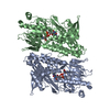
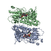
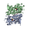
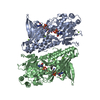
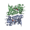
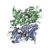
- PDB-4gap: Structure of the Ndi1 protein from Saccharomyces cerevisiae in co... -
+ Open data

- Basic information
| Entry | Database: PDB / ID: 4gap | ||||||
|---|---|---|---|---|---|---|---|
| Title | Structure of the Ndi1 protein from Saccharomyces cerevisiae in complex with NAD+ | ||||||
 Components Components | Rotenone-insensitive NADH-ubiquinone oxidoreductase | ||||||
 Keywords Keywords |  OXIDOREDUCTASE / Nucleotide-binding domains / Membrane OXIDOREDUCTASE / Nucleotide-binding domains / Membrane | ||||||
| Function / homology |  Function and homology information Function and homology informationNADH:quinone reductase (non-electrogenic) / NADH oxidation / mitochondrial electron transport, NADH to ubiquinone / NADH dehydrogenase (ubiquinone) activity / mitochondrial inner membrane / oxidoreductase activity / mitochondrial matrix / positive regulation of apoptotic process / mitochondrion / identical protein bindingSimilarity search - Function | ||||||
| Biological species |  Saccharomyces cerevisiae (brewer's yeast) Saccharomyces cerevisiae (brewer's yeast) | ||||||
| Method | X-RAY DIFFRACTION / SYNCHROTRON / MOLECULAR REPLACEMENT / Resolution: 2.9 Å | ||||||
 Authors Authors | Iwata, M. / Lee, Y. / Yamashita, T. / Yagi, T. / Iwata, S. / Cameron, A.D. / Maher, M.J. | ||||||
 Citation Citation | Journal: Proc.Natl.Acad.Sci.USA / Year: 2012 Title: The structure of the yeast NADH dehydrogenase (Ndi1) reveals overlapping binding sites for water- and lipid-soluble substrates. Authors: Iwata, M. / Lee, Y. / Yamashita, T. / Yagi, T. / Iwata, S. / Cameron, A.D. / Maher, M.J. | ||||||
| History |
|
- Structure visualization
| Structure viewer | Molecule: MolmilJmol/JSmol |
|---|
- Downloads & links
-Download
| PDBx/mmCIF format | 4gap.cif.gz | 381.6 KB | Display | PDBx/mmCIF format |
|---|---|---|---|---|
| PDB format | pdb4gap.ent.gz | 314.9 KB | Display | PDB format |
| PDBx/mmJSON format | 4gap.json.gz | Tree view | PDBx/mmJSON format | |
| Others |  Other downloads Other downloads |
-Validation report
| Arichive directory | https://data.pdbj.org/pub/pdb/validation_reports/ga/4gapftp://data.pdbj.org/pub/pdb/validation_reports/ga/4gap | HTTPS FTP |
|---|
-Related structure data


-Links
 PDBj
PDBj


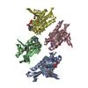
- Assembly
| Deposited unit | 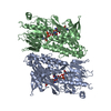
| ||||||||||||||||||
|---|---|---|---|---|---|---|---|---|---|---|---|---|---|---|---|---|---|---|---|
| 1 |
| ||||||||||||||||||
| Unit cell |
| ||||||||||||||||||
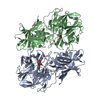
| Noncrystallographic symmetry (NCS) | NCS domain:
NCS domain segments: Component-ID: 1 / Ens-ID: 1 / Beg auth comp-ID: MET / Beg label comp-ID: MET / End auth comp-ID: LEU / End label comp-ID: LEU / Refine code: 1 / Auth seq-ID: 43 - 513 / Label seq-ID: 1 - 471
|
-Components
| #1: Protein | Mass: 52800.562 Da / Num. of mol.: 2 Source method: isolated from a genetically manipulated source Source: (gene. exp.) Saccharomyces cerevisiae (brewer's yeast)Strain: ATCC 204508 / S288c / Gene: NDI1, YML120C, YM7056.06C / Production host:  Escherichia coli (E. coli) Escherichia coli (E. coli)References: UniProt: P32340, NADH:quinone reductase (non-electrogenic) #2: Chemical | Flavin adenine dinucleotide  Mass: 785.550 Da / Num. of mol.: 2 / Source method: obtained synthetically / Formula: C27H33N9O15P2 / Comment: FAD*YM Mass: 785.550 Da / Num. of mol.: 2 / Source method: obtained synthetically / Formula: C27H33N9O15P2 / Comment: FAD*YM#3: Chemical | Nicotinamide adenine dinucleotide  Mass: 663.425 Da / Num. of mol.: 2 / Source method: obtained synthetically / Formula: C21H27N7O14P2 / Comment: NAD*YM Mass: 663.425 Da / Num. of mol.: 2 / Source method: obtained synthetically / Formula: C21H27N7O14P2 / Comment: NAD*YM |
|---|
-Experimental details
-Experiment
| Experiment | Method: X-RAY DIFFRACTION / Number of used crystals: 1 |
|---|
- Sample preparation
| Crystal | Density Matthews: 3.17 Å3/Da / Density % sol: 61.21 % |
|---|---|
| Crystal grow | Temperature: 293 K / Method: vapor diffusion, hanging drop / pH: 6 Details: 50 mM MES, 34% PEG 400, 100 mM NaCl, 2% Ethylene Glycol, 5% glycerol, 5 mM NAD+, pH 6.0, VAPOR DIFFUSION, HANGING DROP, temperature 293K |
-Data collection
| Diffraction | Mean temperature: 100 K |
|---|---|
| Diffraction source | Source: SYNCHROTRON / Site: ESRF  / Beamline: ID29 / Wavelength: 0.98 Å / Beamline: ID29 / Wavelength: 0.98 Å |
| Detector | Type: ADSC QUANTUM 315r / Detector: CCD / Date: Jan 1, 2006 |
| Radiation | Protocol: SINGLE WAVELENGTH / Monochromatic (M) / Laue (L): M / Scattering type: x-ray |
| Radiation wavelength | Wavelength: 0.98 Å / Relative weight: 1 |
| Reflection | Resolution: 2.9→40 Å / Num. all: 29065 / Num. obs: 29065 / % possible obs: 95.1 % / Observed criterion σ(F): 0 / Observed criterion σ(I): 0 |
- Processing
| Software |
| |||||||||||||||||||||||||||||||||||||||||||||||||||||||||||||||||||||||||||
|---|---|---|---|---|---|---|---|---|---|---|---|---|---|---|---|---|---|---|---|---|---|---|---|---|---|---|---|---|---|---|---|---|---|---|---|---|---|---|---|---|---|---|---|---|---|---|---|---|---|---|---|---|---|---|---|---|---|---|---|---|---|---|---|---|---|---|---|---|---|---|---|---|---|---|---|---|
| Refinement | Method to determine structure: MOLECULAR REPLACEMENT / Resolution: 2.9→39.84 Å / Cor.coef. Fo:Fc: 0.912 / Cor.coef. Fo:Fc free: 0.87 / Occupancy max: 1 / Occupancy min: 0.25 / SU B: 36.933 / SU ML: 0.338 / Cross valid method: THROUGHOUT / σ(F): 0 / ESU R Free: 0.435 / Stereochemistry target values: MAXIMUM LIKELIHOOD / Details: HYDROGENS HAVE BEEN ADDED IN THE RIDING POSITIONS
| |||||||||||||||||||||||||||||||||||||||||||||||||||||||||||||||||||||||||||
| Solvent computation | Ion probe radii: 0.8 Å / Shrinkage radii: 0.8 Å / VDW probe radii: 1.2 Å / Solvent model: BABINET MODEL WITH MASK | |||||||||||||||||||||||||||||||||||||||||||||||||||||||||||||||||||||||||||
| Displacement parameters | Biso max: 72.51 Å2 / Biso mean: 63.134 Å2 / Biso min: 14.45 Å2
| |||||||||||||||||||||||||||||||||||||||||||||||||||||||||||||||||||||||||||
| Refinement step | Cycle: LAST / Resolution: 2.9→39.84 Å
| |||||||||||||||||||||||||||||||||||||||||||||||||||||||||||||||||||||||||||
| Refine LS restraints |
| |||||||||||||||||||||||||||||||||||||||||||||||||||||||||||||||||||||||||||
| Refine LS restraints NCS | Number: 6274 / Type: TIGHT THERMAL / Rms dev position: 2.63 Å / Weight position: 0.5 | |||||||||||||||||||||||||||||||||||||||||||||||||||||||||||||||||||||||||||
| LS refinement shell | Resolution: 2.9→2.975 Å / Total num. of bins used: 20
| |||||||||||||||||||||||||||||||||||||||||||||||||||||||||||||||||||||||||||
| Refinement TLS params. | Method: refined / Refine-ID: X-RAY DIFFRACTION
| |||||||||||||||||||||||||||||||||||||||||||||||||||||||||||||||||||||||||||
| Refinement TLS group |
|
