 Movie
Movie Controller

[English] 日本語
 Yorodumi
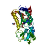
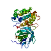
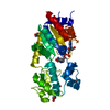
- PDB-4g3g: Crystal structure of murine NF-kappaB inducing kinase (NIK) V408L... -
+ Open data

- Basic information
| Entry | Database: PDB / ID: 4g3g | ||||||
|---|---|---|---|---|---|---|---|
| Title | Crystal structure of murine NF-kappaB inducing kinase (NIK) V408L bound to a 2-(aminothiazolyl)phenol (cmp3) | ||||||
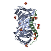
 Components Components | NF-kappa-beta-inducing kinase | ||||||
 Keywords Keywords | TRANSFERASE/TRANSFERASE INHIBITOR / non-RD kinase / protein serine/threonine kinase /  NF-kappaB / MAP3K14 / Structure-based drug design / transferase / TRANSFERASE-TRANSFERASE INHIBITOR complex NF-kappaB / MAP3K14 / Structure-based drug design / transferase / TRANSFERASE-TRANSFERASE INHIBITOR complex | ||||||
| Function / homology |  Function and homology information Function and homology informationCD28 dependent PI3K/Akt signaling / TNF receptor superfamily (TNFSF) members mediating non-canonical NF-kB pathway / Dectin-1 mediated noncanonical NF-kB signaling / NIK-->noncanonical NF-kB signaling / TNFR2 non-canonical NF-kB pathway / mitogen-activated protein kinase kinase kinase / non-canonical NF-kappaB signal transduction / MAP kinase kinase kinase activity / canonical NF-kappaB signal transduction / fibrillar center ...CD28 dependent PI3K/Akt signaling / TNF receptor superfamily (TNFSF) members mediating non-canonical NF-kB pathway / Dectin-1 mediated noncanonical NF-kB signaling / NIK-->noncanonical NF-kB signaling / TNFR2 non-canonical NF-kB pathway / mitogen-activated protein kinase kinase kinase / non-canonical NF-kappaB signal transduction / MAP kinase kinase kinase activity / canonical NF-kappaB signal transduction / fibrillar center / cellular response to mechanical stimulus / defense response to virus / protein kinase activity / immune response / phosphorylation / protein serine kinase activity / intracellular membrane-bounded organelle / protein serine/threonine kinase activity / nucleoplasm / ATP binding / cytosolSimilarity search - Function | ||||||
| Biological species |  Mus musculus (house mouse) Mus musculus (house mouse) | ||||||
| Method | X-RAY DIFFRACTION / SYNCHROTRON / MOLECULAR REPLACEMENT / Resolution: 2.5 Å | ||||||
 Authors Authors | Hymowitz, S. | ||||||
 Citation Citation | Journal: Structure / Year: 2012 Title: The crystal structure of the catalytic domain of the NF-kappaB inducing kinase reveals a narrow but flexible active site. Authors: de Leon-Boenig, G. / Bowman, K.K. / Feng, J.A. / Crawford, T. / Everett, C. / Franke, Y. / Oh, A. / Stanley, M. / Staben, S.T. / Starovasnik, M.A. / Wallweber, H.J. / Wu, J. / Wu, L.C. / ...Authors: de Leon-Boenig, G. / Bowman, K.K. / Feng, J.A. / Crawford, T. / Everett, C. / Franke, Y. / Oh, A. / Stanley, M. / Staben, S.T. / Starovasnik, M.A. / Wallweber, H.J. / Wu, J. / Wu, L.C. / Johnson, A.R. / Hymowitz, S.G. | ||||||
| History |
|
- Structure visualization
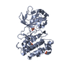
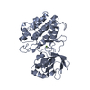
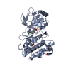
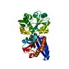
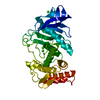
| Structure viewer | Molecule: MolmilJmol/JSmol |
|---|
- Downloads & links
-Download
| PDBx/mmCIF format | 4g3g.cif.gz | 136.9 KB | Display | PDBx/mmCIF format |
|---|---|---|---|---|
| PDB format | pdb4g3g.ent.gz | 107.2 KB | Display | PDB format |
| PDBx/mmJSON format | 4g3g.json.gz | Tree view | PDBx/mmJSON format | |
| Others |  Other downloads Other downloads |
-Validation report
| Arichive directory | https://data.pdbj.org/pub/pdb/validation_reports/g3/4g3gftp://data.pdbj.org/pub/pdb/validation_reports/g3/4g3g | HTTPS FTP |
|---|
-Related structure data


-Links
 PDBj
PDBj
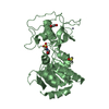
- Assembly
| Deposited unit | 
| ||||||||
|---|---|---|---|---|---|---|---|---|---|
| 1 |
| ||||||||
| Unit cell |
| ||||||||
| Components on special symmetry positions |
|
-Components
| #1: Protein | Mass: 38805.578 Da / Num. of mol.: 1 Source method: isolated from a genetically manipulated source Source: (gene. exp.) Mus musculus (house mouse) / Gene: Map3k14, NF-kappaB inducing kinase (NIK), Nik / Plasmid: pAcGP67 / Production host:   Spodoptera frugiperda (fall armyworm) / Strain (production host): SF9 Spodoptera frugiperda (fall armyworm) / Strain (production host): SF9References: UniProt: Q9WUL6, mitogen-activated protein kinase kinase kinase |
|---|---|
| #2: Chemical | ChemComp-0WA /   Mass: 287.312 Da / Num. of mol.: 1 / Source method: obtained synthetically / Formula: C14H10FN3OS Mass: 287.312 Da / Num. of mol.: 1 / Source method: obtained synthetically / Formula: C14H10FN3OS |
| #3: Water | ChemComp-HOH / Water Mass: 18.015 Da / Num. of mol.: 38 / Source method: isolated from a natural source / Formula: H2O Mass: 18.015 Da / Num. of mol.: 38 / Source method: isolated from a natural source / Formula: H2O |
-Experimental details
-Experiment
| Experiment | Method: X-RAY DIFFRACTION / Number of used crystals: 1 |
|---|
- Sample preparation
| Crystal | Density Matthews: 2.08 Å3/Da / Density % sol: 40.99 % |
|---|---|
| Crystal grow | Temperature: 277 K / Method: vapor diffusion, sitting drop / pH: 7 Details: 0.2 uL protein plus 0.2 uL of 0.5 mM Cmp3, 100 mM HEPES pH 7.0, 0.01 M zinc chloride, 20% PEG 6000, VAPOR DIFFUSION, SITTING DROP, temperature 277K |
-Data collection
| Diffraction | Mean temperature: 93 K |
|---|---|
| Diffraction source | Source: SYNCHROTRON / Site: SSRL  / Beamline: BL7-1 / Wavelength: 0.97946 Å / Beamline: BL7-1 / Wavelength: 0.97946 Å |
| Detector | Type: ADSC QUANTUM 315 / Detector: CCD / Date: Jan 13, 2010 |
| Radiation | Monochromator: Si(111) / Protocol: SINGLE WAVELENGTH / Monochromatic (M) / Laue (L): M / Scattering type: x-ray |
| Radiation wavelength | Wavelength: 0.97946 Å / Relative weight: 1 |
| Reflection | Resolution: 2.5→46.57 Å / Num. all: 11486 / Num. obs: 11486 / % possible obs: 99.3 % / Observed criterion σ(F): 0 / Observed criterion σ(I): -3 / Redundancy: 6.8 % / Rsym value: 0.119 / Net I/σ(I): 15.6 |
| Reflection shell | Resolution: 2.5→2.59 Å / Redundancy: 3.8 % / Mean I/σ(I) obs: 1.8 / Num. unique all: 1051 / Rsym value: 0.619 / % possible all: 93.3 |
- Processing
| Software |
| |||||||||||||||||||||||||||||||||||||||||||||
|---|---|---|---|---|---|---|---|---|---|---|---|---|---|---|---|---|---|---|---|---|---|---|---|---|---|---|---|---|---|---|---|---|---|---|---|---|---|---|---|---|---|---|---|---|---|---|
| Refinement | Method to determine structure: MOLECULAR REPLACEMENT / Resolution: 2.5→46.57 Å / Cor.coef. Fo:Fc: 0.936 / Cor.coef. Fo:Fc free: 0.905 / SU B: 19.01 / SU ML: 0.209 / Cross valid method: THROUGHOUT / σ(I): 0 / ESU R: 1.69 / ESU R Free: 0.323 / Stereochemistry target values: MAXIMUM LIKELIHOOD / Details: HYDROGENS HAVE BEEN USED IF PRESENT IN THE INPUT
| |||||||||||||||||||||||||||||||||||||||||||||
| Solvent computation | Ion probe radii: 0.8 Å / Shrinkage radii: 0.8 Å / VDW probe radii: 1.4 Å / Solvent model: MASK | |||||||||||||||||||||||||||||||||||||||||||||
| Displacement parameters | Biso mean: 39.35 Å2
| |||||||||||||||||||||||||||||||||||||||||||||
| Refinement step | Cycle: LAST / Resolution: 2.5→46.57 Å
| |||||||||||||||||||||||||||||||||||||||||||||
| Refine LS restraints |
| |||||||||||||||||||||||||||||||||||||||||||||
| LS refinement shell | Resolution: 2.5→2.552 Å / Total num. of bins used: 25
| |||||||||||||||||||||||||||||||||||||||||||||
| Refinement TLS params. | Method: refined / Origin x: 18.7916 Å / Origin y: 17.6438 Å / Origin z: 2.4961 Å
|
