 Movie
Movie Controller


+ Open data

- Basic information
| Entry | Database: PDB / ID: 4fu3 | ||||||
|---|---|---|---|---|---|---|---|
| Title | CID of human RPRD1B | ||||||
 Components Components | Regulation of nuclear pre-mRNA domain-containing protein 1B | ||||||
 Keywords Keywords |  TRANSCRIPTION / Structural Genomics Consortium / SGC / domain swapping TRANSCRIPTION / Structural Genomics Consortium / SGC / domain swapping | ||||||
| Function / homology |  Function and homology information Function and homology informationregulation of cell cycle process / RNA polymerase II promoter clearance / mRNA 3'-end processing / transcription preinitiation complex / RNA polymerase II complex binding / RNA polymerase II C-terminal domain binding / RNA polymerase II transcribes snRNA genes / positive regulation of cell population proliferation / positive regulation of transcription by RNA polymerase II / nucleoplasm ...regulation of cell cycle process / RNA polymerase II promoter clearance / mRNA 3'-end processing / transcription preinitiation complex / RNA polymerase II complex binding / RNA polymerase II C-terminal domain binding / RNA polymerase II transcribes snRNA genes / positive regulation of cell population proliferation / positive regulation of transcription by RNA polymerase II / nucleoplasm / identical protein binding / nucleusSimilarity search - Function | ||||||
| Biological species |  Homo sapiens (human) Homo sapiens (human) | ||||||
| Method | X-RAY DIFFRACTION / SYNCHROTRON / MOLECULAR REPLACEMENT / molecular replacement / Resolution: 1.9 Å | ||||||
 Authors Authors | Ni, Z. / Xu, C. / Tempel, W. / El Bakkouri, M. / Loppnau, P. / Guo, X. / Bountra, C. / Arrowsmith, C.H. / Edwards, A.M. / Min, J. ...Ni, Z. / Xu, C. / Tempel, W. / El Bakkouri, M. / Loppnau, P. / Guo, X. / Bountra, C. / Arrowsmith, C.H. / Edwards, A.M. / Min, J. / Greenblatt, J.F. / Structural Genomics Consortium (SGC) | ||||||
 Citation Citation | Journal: TO BE PUBLISHED Title: CID of human RPRD1B Authors: Ni, Z. / Xu, C. / Tempel, W. / El Bakkouri, M. / Loppnau, P. / Guo, X. / Bountra, C. / Arrowsmith, C.H. / Edwards, A.M. / Min, J. / Greenblatt, J.F. | ||||||
| History |
|
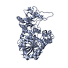
- Structure visualization
| Structure viewer | Molecule: MolmilJmol/JSmol |
|---|
- Downloads & links
-Download
| PDBx/mmCIF format | 4fu3.cif.gz | 116.1 KB | Display | PDBx/mmCIF format |
|---|---|---|---|---|
| PDB format | pdb4fu3.ent.gz | 90.4 KB | Display | PDB format |
| PDBx/mmJSON format | 4fu3.json.gz | Tree view | PDBx/mmJSON format | |
| Others |  Other downloads Other downloads |
-Validation report
| Arichive directory | https://data.pdbj.org/pub/pdb/validation_reports/fu/4fu3ftp://data.pdbj.org/pub/pdb/validation_reports/fu/4fu3 | HTTPS FTP |
|---|
-Related structure data


-Links
 PDBj
PDBj


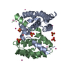
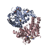
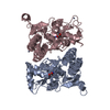
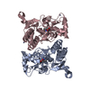
- Assembly
| Deposited unit | 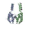
| ||||||||
|---|---|---|---|---|---|---|---|---|---|
| 1 | 
| ||||||||
| 2 | 
| ||||||||
| 3 | 
| ||||||||
| Unit cell |
|
-Components
| #1: Protein | Mass: 15597.543 Da / Num. of mol.: 2 / Fragment: UNP residues 2-235 Source method: isolated from a genetically manipulated source Source: (gene. exp.) Homo sapiens (human) / Gene: RPRD1B, C20orf77, CREPT / Plasmid: pET15 MHL / Production host:  Escherichia coli (E. coli) / Strain (production host): BL21 / References: UniProt: Q9NQG5 Escherichia coli (E. coli) / Strain (production host): BL21 / References: UniProt: Q9NQG5#2: Chemical | Chloride  Mass: 35.453 Da / Num. of mol.: 3 / Source method: obtained synthetically / Formula: Cl Mass: 35.453 Da / Num. of mol.: 3 / Source method: obtained synthetically / Formula: Cl#3: Chemical | ChemComp-UNX /   Num. of mol.: 10 / Source method: obtained synthetically Num. of mol.: 10 / Source method: obtained synthetically#4: Water | ChemComp-HOH / | Water Mass: 18.015 Da / Num. of mol.: 76 / Source method: isolated from a natural source / Formula: H2O Mass: 18.015 Da / Num. of mol.: 76 / Source method: isolated from a natural source / Formula: H2O |
|---|
-Experimental details
-Experiment
| Experiment | Method: X-RAY DIFFRACTION / Number of used crystals: 1 |
|---|
- Sample preparation
| Crystal | Density Matthews: 2.1 Å3/Da / Density % sol: 41.7 % |
|---|---|
| Crystal grow | Temperature: 291 K / Method: vapor diffusion / pH: 8.5 Details: 30% PEG-4000, 0.2M magnesium chloride, 0.1M TRIS hydrochloride, pH 8.5, vapor diffusion, temperature 291K |
-Data collection
| Diffraction |
| |||||||||||||||||||||||||||||||||||||||||||||||||||||||||||||||||||||||||||||
|---|---|---|---|---|---|---|---|---|---|---|---|---|---|---|---|---|---|---|---|---|---|---|---|---|---|---|---|---|---|---|---|---|---|---|---|---|---|---|---|---|---|---|---|---|---|---|---|---|---|---|---|---|---|---|---|---|---|---|---|---|---|---|---|---|---|---|---|---|---|---|---|---|---|---|---|---|---|---|
| Diffraction source | Source: SYNCHROTRON / Site: CHESS  / Beamline: F1 / Wavelength: 0.9179 Å / Beamline: F1 / Wavelength: 0.9179 Å | |||||||||||||||||||||||||||||||||||||||||||||||||||||||||||||||||||||||||||||
| Detector | Type: ADSC QUANTUM 270 / Detector: CCD / Date: Oct 19, 2011 | |||||||||||||||||||||||||||||||||||||||||||||||||||||||||||||||||||||||||||||
| Radiation | Protocol: SINGLE WAVELENGTH / Monochromatic (M) / Laue (L): M / Scattering type: x-ray | |||||||||||||||||||||||||||||||||||||||||||||||||||||||||||||||||||||||||||||
| Radiation wavelength | Wavelength: 0.9179 Å / Relative weight: 1 | |||||||||||||||||||||||||||||||||||||||||||||||||||||||||||||||||||||||||||||
| Reflection | Resolution: 1.9→46.78 Å / Num. obs: 20170 / % possible obs: 99.95 % / Redundancy: 7.19 % / Rmerge(I) obs: 0.07 / Net I/σ(I): 19.6638 | |||||||||||||||||||||||||||||||||||||||||||||||||||||||||||||||||||||||||||||
| Reflection shell |
|
-Phasing
| Phasing | Method: molecular replacement |
|---|
- Processing
| Software |
| ||||||||||||||||||||||||||||||||||||||||||||||||||||||||||||||||||||||||||||||||||||||||||||||||||||||||||||
|---|---|---|---|---|---|---|---|---|---|---|---|---|---|---|---|---|---|---|---|---|---|---|---|---|---|---|---|---|---|---|---|---|---|---|---|---|---|---|---|---|---|---|---|---|---|---|---|---|---|---|---|---|---|---|---|---|---|---|---|---|---|---|---|---|---|---|---|---|---|---|---|---|---|---|---|---|---|---|---|---|---|---|---|---|---|---|---|---|---|---|---|---|---|---|---|---|---|---|---|---|---|---|---|---|---|---|---|---|---|
| Refinement | Method to determine structure: MOLECULAR REPLACEMENT Starting model: unpublished model of same protein, different crystal form Resolution: 1.9→19.77 Å / Cor.coef. Fo:Fc: 0.9456 / Cor.coef. Fo:Fc free: 0.8974 / Occupancy max: 1 / Occupancy min: 0.5 / SU R Cruickshank DPI: 0.17 / Cross valid method: THROUGHOUT / σ(F): 0 Details: DM, PARROT, ARP/WARP, REFMAC, COOT and the MOLPROBITY server were also used.
| ||||||||||||||||||||||||||||||||||||||||||||||||||||||||||||||||||||||||||||||||||||||||||||||||||||||||||||
| Displacement parameters | Biso max: 107.05 Å2 / Biso mean: 37.5815 Å2 / Biso min: 16.61 Å2
| ||||||||||||||||||||||||||||||||||||||||||||||||||||||||||||||||||||||||||||||||||||||||||||||||||||||||||||
| Refine analyze | Luzzati coordinate error obs: 0.301 Å | ||||||||||||||||||||||||||||||||||||||||||||||||||||||||||||||||||||||||||||||||||||||||||||||||||||||||||||
| Refinement step | Cycle: LAST / Resolution: 1.9→19.77 Å
| ||||||||||||||||||||||||||||||||||||||||||||||||||||||||||||||||||||||||||||||||||||||||||||||||||||||||||||
| Refine LS restraints |
| ||||||||||||||||||||||||||||||||||||||||||||||||||||||||||||||||||||||||||||||||||||||||||||||||||||||||||||
| LS refinement shell | Resolution: 1.9→2 Å / Total num. of bins used: 10
| ||||||||||||||||||||||||||||||||||||||||||||||||||||||||||||||||||||||||||||||||||||||||||||||||||||||||||||
| Refinement TLS params. | Method: refined / Refine-ID: X-RAY DIFFRACTION
| ||||||||||||||||||||||||||||||||||||||||||||||||||||||||||||||||||||||||||||||||||||||||||||||||||||||||||||
| Refinement TLS group |
|
