 Movie
Movie Controller


+ Open data

- Basic information
| Entry | Database: PDB / ID: 4frx | ||||||
|---|---|---|---|---|---|---|---|
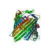
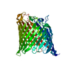
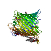
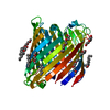
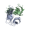
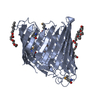
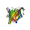
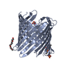
| Title | Crystal Structure of Pseudomonas aeruginosa OccK8 (OprE) | ||||||
 Components Components | Anaerobically-induced outer membrane porin OprE | ||||||
 Keywords Keywords |  TRANSPORT PROTEIN / beta-barrel / transporter (porin) / outer membrane TRANSPORT PROTEIN / beta-barrel / transporter (porin) / outer membrane | ||||||
| Function / homology |  Function and homology information Function and homology informationouter membrane / channel activity / porin activity / membrane => GO:0016020Similarity search - Function | ||||||
| Biological species |   Pseudomonas aeruginosa (bacteria) Pseudomonas aeruginosa (bacteria) | ||||||
| Method | X-RAY DIFFRACTION / SYNCHROTRON / MOLECULAR REPLACEMENT / Resolution: 1.9 Å | ||||||
 Authors Authors | Eren, E. / van den Berg, B. | ||||||
 Citation Citation | Journal: To be Published Title: Crystal Structures of OccK Subfamily Proteins Authors: Eren, E. / van den Berg, B. | ||||||
| History |
|
- Structure visualization
| Structure viewer | Molecule: MolmilJmol/JSmol |
|---|
- Downloads & links
-Download
| PDBx/mmCIF format | 4frx.cif.gz | 326.8 KB | Display | PDBx/mmCIF format |
|---|---|---|---|---|
| PDB format | pdb4frx.ent.gz | 266 KB | Display | PDB format |
| PDBx/mmJSON format | 4frx.json.gz | Tree view | PDBx/mmJSON format | |
| Others |  Other downloads Other downloads |
-Validation report
| Arichive directory | https://data.pdbj.org/pub/pdb/validation_reports/fr/4frxftp://data.pdbj.org/pub/pdb/validation_reports/fr/4frx | HTTPS FTP |
|---|
-Related structure data
| Related structure data |  4frtC  4fsoC  4fspC  4ft6C 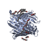 3sysS S: Starting model for refinement C: citing same article ( |
|---|---|
| Similar structure data |


-Links
 PDBj
PDBj- Assembly
| Deposited unit | 
| |||||||||
|---|---|---|---|---|---|---|---|---|---|---|
| 1 | 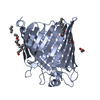
| |||||||||
| 2 | 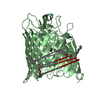
| |||||||||
| Unit cell |
| |||||||||
| Components on special symmetry positions |
|
-Components
| #1: Protein | Mass: 47579.074 Da / Num. of mol.: 2 Source method: isolated from a genetically manipulated source Source: (gene. exp.) Pseudomonas aeruginosa (bacteria) / Strain: ATCC 15692 / PAO1 / 1C / PRS 101 / LMG 12228 / Gene: oprE, PA0291 / Production host: Escherichia coli (E. coli) / References: UniProt: G3XDA5#2: Chemical | ChemComp-C8E / (   Mass: 306.438 Da / Num. of mol.: 9 / Source method: obtained synthetically / Formula: C16H34O5 / Comment: C8E, detergent*YM Mass: 306.438 Da / Num. of mol.: 9 / Source method: obtained synthetically / Formula: C16H34O5 / Comment: C8E, detergent*YM#3: Water | ChemComp-HOH / | Water Mass: 18.015 Da / Num. of mol.: 574 / Source method: isolated from a natural source / Formula: H2O Mass: 18.015 Da / Num. of mol.: 574 / Source method: isolated from a natural source / Formula: H2O |
|---|
-Experimental details
-Experiment
| Experiment | Method: X-RAY DIFFRACTION / Number of used crystals: 1 |
|---|
- Sample preparation
| Crystal | Density Matthews: 3.39 Å3/Da / Density % sol: 63.73 % |
|---|---|
| Crystal grow | Temperature: 293 K / Method: vapor diffusion, hanging drop / pH: 7 Details: 25% PEG 400, 0.2M ammonium sulphate, 0.05M HEPES, pH 7.0, VAPOR DIFFUSION, HANGING DROP, temperature 293K |
-Data collection
| Diffraction | Mean temperature: 100 K |
|---|---|
| Diffraction source | Source: SYNCHROTRON / Site: NSLS  / Beamline: X25 / Wavelength: 1.1 Å / Beamline: X25 / Wavelength: 1.1 Å |
| Detector | Type: ADSC QUANTUM 315 / Detector: CCD / Date: Feb 1, 2012 |
| Radiation | Monochromator: Double silicon(111) crystal monochromator / Protocol: SINGLE WAVELENGTH / Monochromatic (M) / Laue (L): M / Scattering type: x-ray |
| Radiation wavelength | Wavelength: 1.1 Å / Relative weight: 1 |
| Reflection | Resolution: 1.9→50 Å / Num. obs: 99602 / % possible obs: 99.3 % / Redundancy: 3.5 % / Rsym value: 0.118 / Net I/σ(I): 9.2 |
| Reflection shell | Resolution: 1.9→1.93 Å / Redundancy: 3.4 % / Mean I/σ(I) obs: 1.93 / Num. unique all: 4913 / Rsym value: 0.553 / % possible all: 98.6 |
- Processing
| Software |
| |||||||||||||||||||||||||||||||||||||||||||||||||||||||||||||||||||||||||||||||||||||||||||||||||||||||||
|---|---|---|---|---|---|---|---|---|---|---|---|---|---|---|---|---|---|---|---|---|---|---|---|---|---|---|---|---|---|---|---|---|---|---|---|---|---|---|---|---|---|---|---|---|---|---|---|---|---|---|---|---|---|---|---|---|---|---|---|---|---|---|---|---|---|---|---|---|---|---|---|---|---|---|---|---|---|---|---|---|---|---|---|---|---|---|---|---|---|---|---|---|---|---|---|---|---|---|---|---|---|---|---|---|---|---|
| Refinement | Method to determine structure: MOLECULAR REPLACEMENT Starting model: PDB ENTRY 3SYS Resolution: 1.9→14.993 Å / SU ML: 0.21 / σ(F): 1.35 / Phase error: 22.44 / Stereochemistry target values: ML
| |||||||||||||||||||||||||||||||||||||||||||||||||||||||||||||||||||||||||||||||||||||||||||||||||||||||||
| Solvent computation | Shrinkage radii: 0.6 Å / VDW probe radii: 0.9 Å / Solvent model: FLAT BULK SOLVENT MODEL / Bsol: 54.336 Å2 / ksol: 0.44 e/Å3 | |||||||||||||||||||||||||||||||||||||||||||||||||||||||||||||||||||||||||||||||||||||||||||||||||||||||||
| Displacement parameters |
| |||||||||||||||||||||||||||||||||||||||||||||||||||||||||||||||||||||||||||||||||||||||||||||||||||||||||
| Refinement step | Cycle: LAST / Resolution: 1.9→14.993 Å
| |||||||||||||||||||||||||||||||||||||||||||||||||||||||||||||||||||||||||||||||||||||||||||||||||||||||||
| Refine LS restraints |
| |||||||||||||||||||||||||||||||||||||||||||||||||||||||||||||||||||||||||||||||||||||||||||||||||||||||||
| LS refinement shell |
| |||||||||||||||||||||||||||||||||||||||||||||||||||||||||||||||||||||||||||||||||||||||||||||||||||||||||
| Refinement TLS params. | Method: refined / Origin x: 31.2622 Å / Origin y: -6.8936 Å / Origin z: 74.2133 Å
| |||||||||||||||||||||||||||||||||||||||||||||||||||||||||||||||||||||||||||||||||||||||||||||||||||||||||
| Refinement TLS group | Selection details: all |
