 Movie
Movie Controller

[English] 日本語
 Yorodumi
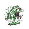
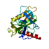
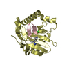
- PDB-4fpw: Crystal Structure of CalU16 from Micromonospora echinospora. Nort... -
+ Open data

- Basic information
| Entry | Database: PDB / ID: 4fpw | ||||||
|---|---|---|---|---|---|---|---|
| Title | Crystal Structure of CalU16 from Micromonospora echinospora. Northeast Structural Genomics Consortium Target MiR12. | ||||||
 Components Components | CalU16 | ||||||
 Keywords Keywords |  Structural Genomics / Unknown Function / PSI-Biology / Northeast Structural Genomics Consortium / NESG / CalU16 Structural Genomics / Unknown Function / PSI-Biology / Northeast Structural Genomics Consortium / NESG / CalU16 | ||||||
| Function / homology | Activator of Hsp90 ATPase homologue 1-like / Activator of Hsp90 ATPase homolog 1-like protein / START domain / Alpha-D-Glucose-1,6-Bisphosphate; Chain A, domain 4 / START-like domain superfamily / 2-Layer Sandwich / Alpha Beta / CalU16 Function and homology information Function and homology information | ||||||
| Biological species |  Micromonospora echinospora (bacteria) Micromonospora echinospora (bacteria) | ||||||
| Method | X-RAY DIFFRACTION / SYNCHROTRON / SAD / Resolution: 2.5 Å | ||||||
 Authors Authors | Seetharaman, J. / Lew, S. / Janjua, H. / Xiao, R. / Acton, T.B. / Everett, J.K. / Phillips Jr., G.N. / Kennedy, M.A. / Montelione, G.T. / Hunt, J.F. ...Seetharaman, J. / Lew, S. / Janjua, H. / Xiao, R. / Acton, T.B. / Everett, J.K. / Phillips Jr., G.N. / Kennedy, M.A. / Montelione, G.T. / Hunt, J.F. / Tong, L. / Northeast Structural Genomics Consortium (NESG) | ||||||
 Citation Citation | Journal: Acs Chem.Biol. / Year: 2014 Title: Structure-Guided Functional Characterization of Enediyne Self-Sacrifice Resistance Proteins, CalU16 and CalU19. Authors: Elshahawi, S.I. / Ramelot, T.A. / Seetharaman, J. / Chen, J. / Singh, S. / Yang, Y. / Pederson, K. / Kharel, M.K. / Xiao, R. / Lew, S. / Yennamalli, R.M. / Miller, M.D. / Wang, F. / Tong, L. ...Authors: Elshahawi, S.I. / Ramelot, T.A. / Seetharaman, J. / Chen, J. / Singh, S. / Yang, Y. / Pederson, K. / Kharel, M.K. / Xiao, R. / Lew, S. / Yennamalli, R.M. / Miller, M.D. / Wang, F. / Tong, L. / Montelione, G.T. / Kennedy, M.A. / Bingman, C.A. / Zhu, H. / Phillips, G.N. / Thorson, J.S. | ||||||
| History |
|
- Structure visualization
| Structure viewer | Molecule: MolmilJmol/JSmol |
|---|
- Downloads & links
-Download
| PDBx/mmCIF format | 4fpw.cif.gz | 135.5 KB | Display | PDBx/mmCIF format |
|---|---|---|---|---|
| PDB format | pdb4fpw.ent.gz | 113.1 KB | Display | PDB format |
| PDBx/mmJSON format | 4fpw.json.gz | Tree view | PDBx/mmJSON format | |
| Others |  Other downloads Other downloads |
-Validation report
| Arichive directory | https://data.pdbj.org/pub/pdb/validation_reports/fp/4fpwftp://data.pdbj.org/pub/pdb/validation_reports/fp/4fpw | HTTPS FTP |
|---|
-Related structure data


-Links
 PDBj
PDBj- Assembly
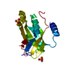
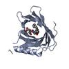
| Deposited unit | 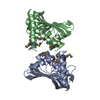
| |||||||||||||||||||||||||||
|---|---|---|---|---|---|---|---|---|---|---|---|---|---|---|---|---|---|---|---|---|---|---|---|---|---|---|---|---|
| 1 | 
| |||||||||||||||||||||||||||
| 2 | 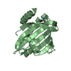
| |||||||||||||||||||||||||||
| 3 |
| |||||||||||||||||||||||||||
| Unit cell |
| |||||||||||||||||||||||||||
| Noncrystallographic symmetry (NCS) | NCS domain:
NCS domain segments:
|
-Components
| #1: Protein | Mass: 20235.729 Da / Num. of mol.: 2 Source method: isolated from a genetically manipulated source Source: (gene. exp.) Micromonospora echinospora (bacteria) / Gene: calU16 / Production host: Escherichia coli (E. coli) / References: UniProt: Q8KNE9#2: Water | ChemComp-HOH / | Water Mass: 18.015 Da / Num. of mol.: 128 / Source method: isolated from a natural source / Formula: H2O Mass: 18.015 Da / Num. of mol.: 128 / Source method: isolated from a natural source / Formula: H2O |
|---|
-Experimental details
-Experiment
| Experiment | Method: X-RAY DIFFRACTION / Number of used crystals: 1 |
|---|
- Sample preparation
| Crystal | Density Matthews: 2.95 Å3/Da / Density % sol: 58.31 % |
|---|---|
| Crystal grow | Temperature: 291 K / Method: microbatch under oil / pH: 7 Details: 2.9 M Sodium malonate pH 7, Microbatch under oil, temperature 291K |
-Data collection
| Diffraction |
| |||||||||||||||
|---|---|---|---|---|---|---|---|---|---|---|---|---|---|---|---|---|
| Diffraction source |
| |||||||||||||||
| Detector |
| |||||||||||||||
| Radiation | Monochromator: KOHZU double crystal monochromator / Protocol: SINGLE WAVELENGTH / Monochromatic (M) / Laue (L): M / Scattering type: x-ray | |||||||||||||||
| Radiation wavelength | Wavelength: 0.979 Å / Relative weight: 1 | |||||||||||||||
| Reflection twin |
| |||||||||||||||
| Reflection | Resolution: 2.5→50 Å / Num. all: 21977 / Num. obs: 21977 / % possible obs: 94.2 % / Observed criterion σ(F): 0 / Observed criterion σ(I): 0 / Redundancy: 6 % / Rmerge(I) obs: 0.063 / Rsym value: 0.057 / Net I/σ(I): 13.7 | |||||||||||||||
| Reflection shell | Resolution: 2.5→2.54 Å / Rmerge(I) obs: 0.225 / % possible all: 99.4 |

- Processing
| Software |
| ||||||||||||||||||||||||||||||||||||||||||||||||||||||||||||||||||||||||||||||||||||||||||||||||||||||||||||||||||||||||||||||||||||||||||||||||||||||||||||||||||||||||||
|---|---|---|---|---|---|---|---|---|---|---|---|---|---|---|---|---|---|---|---|---|---|---|---|---|---|---|---|---|---|---|---|---|---|---|---|---|---|---|---|---|---|---|---|---|---|---|---|---|---|---|---|---|---|---|---|---|---|---|---|---|---|---|---|---|---|---|---|---|---|---|---|---|---|---|---|---|---|---|---|---|---|---|---|---|---|---|---|---|---|---|---|---|---|---|---|---|---|---|---|---|---|---|---|---|---|---|---|---|---|---|---|---|---|---|---|---|---|---|---|---|---|---|---|---|---|---|---|---|---|---|---|---|---|---|---|---|---|---|---|---|---|---|---|---|---|---|---|---|---|---|---|---|---|---|---|---|---|---|---|---|---|---|---|---|---|---|---|---|---|---|---|
| Refinement | Method to determine structure: SAD / Resolution: 2.5→44.91 Å / Cor.coef. Fo:Fc: 0.929 / Cor.coef. Fo:Fc free: 0.902 / SU B: 18.134 / SU ML: 0.217 / Cross valid method: THROUGHOUT / ESU R: 0.098 / ESU R Free: 0.063 / Stereochemistry target values: MAXIMUM LIKELIHOOD / Details: HYDROGENS HAVE BEEN ADDED IN THE RIDING POSITIONS
| ||||||||||||||||||||||||||||||||||||||||||||||||||||||||||||||||||||||||||||||||||||||||||||||||||||||||||||||||||||||||||||||||||||||||||||||||||||||||||||||||||||||||||
| Solvent computation | Ion probe radii: 0.8 Å / Shrinkage radii: 0.8 Å / VDW probe radii: 1.2 Å / Solvent model: MASK | ||||||||||||||||||||||||||||||||||||||||||||||||||||||||||||||||||||||||||||||||||||||||||||||||||||||||||||||||||||||||||||||||||||||||||||||||||||||||||||||||||||||||||
| Displacement parameters | Biso mean: 53.644 Å2
| ||||||||||||||||||||||||||||||||||||||||||||||||||||||||||||||||||||||||||||||||||||||||||||||||||||||||||||||||||||||||||||||||||||||||||||||||||||||||||||||||||||||||||
| Refinement step | Cycle: LAST / Resolution: 2.5→44.91 Å
| ||||||||||||||||||||||||||||||||||||||||||||||||||||||||||||||||||||||||||||||||||||||||||||||||||||||||||||||||||||||||||||||||||||||||||||||||||||||||||||||||||||||||||
| Refine LS restraints |
| ||||||||||||||||||||||||||||||||||||||||||||||||||||||||||||||||||||||||||||||||||||||||||||||||||||||||||||||||||||||||||||||||||||||||||||||||||||||||||||||||||||||||||
| Refine LS restraints NCS | Ens-ID: 1 / Number: 149 / Refine-ID: X-RAY DIFFRACTION / Type: interatomic distance / Rms dev position: 0.24 Å / Weight position: 0.05
| ||||||||||||||||||||||||||||||||||||||||||||||||||||||||||||||||||||||||||||||||||||||||||||||||||||||||||||||||||||||||||||||||||||||||||||||||||||||||||||||||||||||||||
| LS refinement shell | Resolution: 2.501→2.566 Å / Total num. of bins used: 20
| ||||||||||||||||||||||||||||||||||||||||||||||||||||||||||||||||||||||||||||||||||||||||||||||||||||||||||||||||||||||||||||||||||||||||||||||||||||||||||||||||||||||||||
| Refinement TLS params. | Method: refined / Refine-ID: X-RAY DIFFRACTION
| ||||||||||||||||||||||||||||||||||||||||||||||||||||||||||||||||||||||||||||||||||||||||||||||||||||||||||||||||||||||||||||||||||||||||||||||||||||||||||||||||||||||||||
| Refinement TLS group |
|
