 Movie
Movie Controller

[English] 日本語
 Yorodumi
- PDB-4fo1: Crystal structure of lincosamide antibiotic adenylyltransferase L... -
+ Open data

- Basic information
| Entry | Database: PDB / ID: 4fo1 | |||||||||
|---|---|---|---|---|---|---|---|---|---|---|
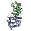
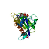
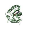
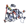
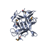
| Title | Crystal structure of lincosamide antibiotic adenylyltransferase LnuA, apo | |||||||||
 Components Components | Lincosamide resistance protein | |||||||||
 Keywords Keywords |  TRANSFERASE / STRUCTURAL GENOMICS / ANTIBIOTIC RESISTANCE / CENTER FOR STRUCTURAL GENOMICS OF INFECTIOUS DISEASES (CSGID) / NIAID / National Institute of Allergy and Infectious Diseases / ALPHA+BETA STRUCTURE / AMINOGLYCOSIDE-2''-ADENYLYLTRANSFERASE SUPERFAMILY / NUCLEOTIDYLTRANSFERASE SUPERFAMILY / LINCOSAMIDE ADENYLYLTRANSFERASE / LINCOSAMIDE ANTIBIOTICS / LINCOMYCIN / CLINDAMYCIN / ADENOSINE TRIPHOSPHATE / INTRACELLULAR TRANSFERASE / STRUCTURAL GENOMICS / ANTIBIOTIC RESISTANCE / CENTER FOR STRUCTURAL GENOMICS OF INFECTIOUS DISEASES (CSGID) / NIAID / National Institute of Allergy and Infectious Diseases / ALPHA+BETA STRUCTURE / AMINOGLYCOSIDE-2''-ADENYLYLTRANSFERASE SUPERFAMILY / NUCLEOTIDYLTRANSFERASE SUPERFAMILY / LINCOSAMIDE ADENYLYLTRANSFERASE / LINCOSAMIDE ANTIBIOTICS / LINCOMYCIN / CLINDAMYCIN / ADENOSINE TRIPHOSPHATE / INTRACELLULAR | |||||||||
| Function / homology | Aminoglycoside-2''-adenylyltransferase / Aminoglycoside-2''-adenylyltransferase / Beta Polymerase; domain 2 - #40 / Beta Polymerase; domain 2 / Nucleotidyltransferase superfamily / response to antibiotic / 2-Layer Sandwich / Alpha Beta / Lincosamide resistance protein Function and homology information Function and homology information | |||||||||
| Biological species |  Staphylococcus haemolyticus (bacteria) Staphylococcus haemolyticus (bacteria) | |||||||||
| Method | X-RAY DIFFRACTION / SYNCHROTRON / SAD / Resolution: 2.15 Å | |||||||||
 Authors Authors | Stogios, P.J. / Wawrzak, Z. / Minasov, G. / Evdokimova, E. / Egorova, O. / Kudritska, M. / Yim, V. / Courvalin, P. / Savchenko, A. / Anderson, W.F. / Center for Structural Genomics of Infectious Diseases (CSGID) | |||||||||
 Citation Citation | Journal: TO BE PUBLISHED Title: Crystal structure of lincosamide antibiotic adenylyltransferase LnuA, apo Authors: Stogios, P.J. / Wawrzak, Z. / Minasov, G. / Evdokimova, E. / Egorova, O. / Kudritska, M. / Yim, V. / Courvalin, P. / Savchenko, A. / Anderson, W.F. / Center for Structural Genomics of Infectious Diseases (CSGID) | |||||||||
| History |
|
- Structure visualization
| Structure viewer | Molecule: MolmilJmol/JSmol |
|---|
- Downloads & links
-Download
| PDBx/mmCIF format | 4fo1.cif.gz | 148.9 KB | Display | PDBx/mmCIF format |
|---|---|---|---|---|
| PDB format | pdb4fo1.ent.gz | 124.2 KB | Display | PDB format |
| PDBx/mmJSON format | 4fo1.json.gz | Tree view | PDBx/mmJSON format | |
| Others |  Other downloads Other downloads |
-Validation report
| Arichive directory | https://data.pdbj.org/pub/pdb/validation_reports/fo/4fo1ftp://data.pdbj.org/pub/pdb/validation_reports/fo/4fo1 | HTTPS FTP |
|---|
-Related structure data
| Related structure data | |
|---|---|
| Similar structure data | |
| Other databases |


-Links
 PDBj
PDBj
- Assembly
| Deposited unit | 
| ||||||||
|---|---|---|---|---|---|---|---|---|---|
| 1 | 
| ||||||||
| 2 | 
| ||||||||
| Unit cell |
|
-Components
| #1: Protein | Mass: 19253.844 Da / Num. of mol.: 2 / Fragment: LnuA Source method: isolated from a genetically manipulated source Source: (gene. exp.) Staphylococcus haemolyticus (bacteria) / Gene: linA / Plasmid: P15TV LIC / Production host: Escherichia coli (E. coli) / Strain (production host): BL21(DE3) / References: UniProt: P06107#2: Chemical | ChemComp-EPE / HEPES  Mass: 238.305 Da / Num. of mol.: 4 / Source method: obtained synthetically / Formula: C8H18N2O4S / Comment: pH buffer*YM Mass: 238.305 Da / Num. of mol.: 4 / Source method: obtained synthetically / Formula: C8H18N2O4S / Comment: pH buffer*YM#3: Water | ChemComp-HOH / | Water Mass: 18.015 Da / Num. of mol.: 223 / Source method: isolated from a natural source / Formula: H2O Mass: 18.015 Da / Num. of mol.: 223 / Source method: isolated from a natural source / Formula: H2O |
|---|
-Experimental details
-Experiment
| Experiment | Method: X-RAY DIFFRACTION / Number of used crystals: 1 |
|---|
- Sample preparation
| Crystal | Density Matthews: 2.75 Å3/Da / Density % sol: 55.35 % |
|---|---|
| Crystal grow | Temperature: 298 K / Method: vapor diffusion, sitting drop / pH: 7.3 Details: 1.4 M SODIUM CITRATE, 0.1 M HEPES PH 7.3, VAPOR DIFFUSION, SITTING DROP, temperature 298K |
-Data collection
| Diffraction | Mean temperature: 100 K |
|---|---|
| Diffraction source | Source: SYNCHROTRON / Site: APS  / Beamline: 21-ID-F / Wavelength: 0.97872 Å / Beamline: 21-ID-F / Wavelength: 0.97872 Å |
| Detector | Type: MARMOSAIC 225 mm CCD / Detector: CCD / Date: Jun 16, 2011 / Details: MIRRORS |
| Radiation | Monochromator: SI-111 CHANNEL / Protocol: SINGLE WAVELENGTH / Monochromatic (M) / Laue (L): M / Scattering type: x-ray |
| Radiation wavelength | Wavelength: 0.97872 Å / Relative weight: 1 |
| Reflection | Resolution: 2→29.855 Å / Num. obs: 28239 / % possible obs: 100 % / Observed criterion σ(F): 0 / Observed criterion σ(I): -2 / Redundancy: 7.7 % / Rsym value: 0.129 / Net I/σ(I): 15.76 |
| Reflection shell | Resolution: 2→2.03 Å / Redundancy: 7.7 % / Mean I/σ(I) obs: 3.75 / Rsym value: 0.569 / % possible all: 100 |
- Processing
| Software |
| |||||||||||||||||||||||||||||||||||||||||||||||||||||||||||||||||||||||||||||||||||||||||||||||||||||||||||||||||||||||||||||
|---|---|---|---|---|---|---|---|---|---|---|---|---|---|---|---|---|---|---|---|---|---|---|---|---|---|---|---|---|---|---|---|---|---|---|---|---|---|---|---|---|---|---|---|---|---|---|---|---|---|---|---|---|---|---|---|---|---|---|---|---|---|---|---|---|---|---|---|---|---|---|---|---|---|---|---|---|---|---|---|---|---|---|---|---|---|---|---|---|---|---|---|---|---|---|---|---|---|---|---|---|---|---|---|---|---|---|---|---|---|---|---|---|---|---|---|---|---|---|---|---|---|---|---|---|---|---|
| Refinement | Method to determine structure: SAD / Resolution: 2.15→29.855 Å / SU ML: 0.11 / Cross valid method: THROUGHOUT / σ(F): 0 / Phase error: 27.53 / Stereochemistry target values: ML
| |||||||||||||||||||||||||||||||||||||||||||||||||||||||||||||||||||||||||||||||||||||||||||||||||||||||||||||||||||||||||||||
| Solvent computation | Shrinkage radii: 0.8 Å / VDW probe radii: 1.1 Å / Solvent model: FLAT BULK SOLVENT MODEL | |||||||||||||||||||||||||||||||||||||||||||||||||||||||||||||||||||||||||||||||||||||||||||||||||||||||||||||||||||||||||||||
| Refinement step | Cycle: LAST / Resolution: 2.15→29.855 Å
| |||||||||||||||||||||||||||||||||||||||||||||||||||||||||||||||||||||||||||||||||||||||||||||||||||||||||||||||||||||||||||||
| Refine LS restraints |
| |||||||||||||||||||||||||||||||||||||||||||||||||||||||||||||||||||||||||||||||||||||||||||||||||||||||||||||||||||||||||||||
| LS refinement shell |
| |||||||||||||||||||||||||||||||||||||||||||||||||||||||||||||||||||||||||||||||||||||||||||||||||||||||||||||||||||||||||||||
| Refinement TLS params. | Method: refined / Refine-ID: X-RAY DIFFRACTION
| |||||||||||||||||||||||||||||||||||||||||||||||||||||||||||||||||||||||||||||||||||||||||||||||||||||||||||||||||||||||||||||
| Refinement TLS group |
|
