 Movie
Movie Controller

[English] 日本語
 Yorodumi
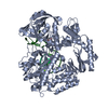
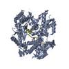
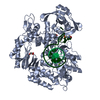
- PDB-4fm1: Pyrococcus abyssi B family DNA polymerase bound to a dsDNA, in ed... -
+ Open data

- Basic information
| Entry | Database: PDB / ID: 4fm1 | ||||||
|---|---|---|---|---|---|---|---|
| Title | Pyrococcus abyssi B family DNA polymerase bound to a dsDNA, in edition mode | ||||||
 Components Components |
| ||||||
 Keywords Keywords | TRANSFERASE/DNA /  DNA polymerase / DNA binding / TRANSFERASE-DNA complex DNA polymerase / DNA binding / TRANSFERASE-DNA complex | ||||||
| Function / homology |  Function and homology information Function and homology informationnucleotide-excision repair, DNA gap filling / DNA replication proofreading / 3'-5'-DNA exonuclease activity / SOS response / base-excision repair, gap-filling / DNA-directed DNA polymerase / DNA-directed DNA polymerase activity / nucleotide binding / DNA bindingSimilarity search - Function | ||||||
| Biological species |   Pyrococcus abyssi (archaea) Pyrococcus abyssi (archaea)synthetic construct (others) | ||||||
| Method | X-RAY DIFFRACTION / SYNCHROTRON / MOLECULAR REPLACEMENT / Resolution: 3 Å | ||||||
 Authors Authors | Gouge, J. / Delarue, M. | ||||||
 Citation Citation | Journal: J.Mol.Biol. / Year: 2012 Title: Molecular Recognition of Canonical and Deaminated Bases by P. abyssi Family B DNA Polymerase. Authors: Gouge, J. / Ralec, C. / Henneke, G. / Delarue, M. | ||||||
| History |
|
- Structure visualization
| Structure viewer | Molecule: MolmilJmol/JSmol |
|---|
- Downloads & links
-Download
| PDBx/mmCIF format | 4fm1.cif.gz | 348.2 KB | Display | PDBx/mmCIF format |
|---|---|---|---|---|
| PDB format | pdb4fm1.ent.gz | 274.7 KB | Display | PDB format |
| PDBx/mmJSON format | 4fm1.json.gz | Tree view | PDBx/mmJSON format | |
| Others |  Other downloads Other downloads |
-Validation report
| Arichive directory | https://data.pdbj.org/pub/pdb/validation_reports/fm/4fm1ftp://data.pdbj.org/pub/pdb/validation_reports/fm/4fm1 | HTTPS FTP |
|---|
-Related structure data
| Related structure data | 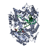 4fltC 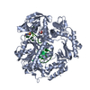 4fluC 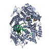 4flvC  4flwSC  4flxC  4flyC  4flzC 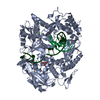 4fm0C  4fm2C C: citing same article ( S: Starting model for refinement |
|---|---|
| Similar structure data |


-Links
 PDBj
PDBj


- Assembly
| Deposited unit | 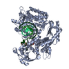
| ||||||||
|---|---|---|---|---|---|---|---|---|---|
| 1 |
| ||||||||
| Unit cell |
|
-Components
-Protein , 1 types, 1 molecules A
| #1: Protein | DNA polymerase I / Pab polymerase Mass: 91809.938 Da / Num. of mol.: 1 / Mutation: D215A Source method: isolated from a genetically manipulated source Source: (gene. exp.) Pyrococcus abyssi (archaea) / Strain: GE5 / Orsay / Gene: polI, pol, PYRAB17200, PAB1128 / Production host:  Escherichia coli (E. coli) / References: UniProt: P0CL77, DNA-directed DNA polymerase Escherichia coli (E. coli) / References: UniProt: P0CL77, DNA-directed DNA polymerase |
|---|
-DNA chain , 2 types, 2 molecules PT
| #2: DNA chain | Mass: 2411.606 Da / Num. of mol.: 1 / Source method: obtained synthetically / Details: chemical synthesis / Source: (synth.) synthetic construct (others) |
|---|---|
| #3: DNA chain | Transcription (biology) Mass: 4008.594 Da / Num. of mol.: 1 / Source method: obtained synthetically / Details: chemical synthesis / Source: (synth.) synthetic construct (others) |
-Non-polymers , 4 types, 260 molecules 


| #4: Chemical | ChemComp-GOL / Glycerol Mass: 92.094 Da / Num. of mol.: 1 / Source method: obtained synthetically / Formula: C3H8O3 Mass: 92.094 Da / Num. of mol.: 1 / Source method: obtained synthetically / Formula: C3H8O3 | ||
|---|---|---|---|
| #5: Chemical | ChemComp-MES / MES (buffer) Mass: 195.237 Da / Num. of mol.: 1 / Source method: obtained synthetically / Formula: C6H13NO4S / Comment: pH buffer*YM Mass: 195.237 Da / Num. of mol.: 1 / Source method: obtained synthetically / Formula: C6H13NO4S / Comment: pH buffer*YM | ||
| #6: Chemical |  Mass: 54.938 Da / Num. of mol.: 2 / Source method: obtained synthetically / Formula: Mn Mass: 54.938 Da / Num. of mol.: 2 / Source method: obtained synthetically / Formula: Mn#7: Water | ChemComp-HOH / | WaterMass: 18.015 Da / Num. of mol.: 256 / Source method: isolated from a natural source / Formula: H2O |
-Experimental details
-Experiment
| Experiment | Method: X-RAY DIFFRACTION / Number of used crystals: 1 |
|---|
- Sample preparation
| Crystal | Density Matthews: 2.57 Å3/Da / Density % sol: 52.21 % |
|---|---|
| Crystal grow | Temperature: 293 K / Method: vapor diffusion, hanging drop / pH: 6.5 Details: 7-12% PEG 20000, 100 mM MES pH 6.5 , VAPOR DIFFUSION, HANGING DROP, temperature 293K |
-Data collection
| Diffraction | Mean temperature: 100 K |
|---|---|
| Diffraction source | Source: SYNCHROTRON / Site: SOLEIL  / Beamline: PROXIMA 1 / Wavelength: 1.89231 Å / Beamline: PROXIMA 1 / Wavelength: 1.89231 Å |
| Detector | Type: ADSC QUANTUM 315r / Detector: CCD / Date: Oct 7, 2010 |
| Radiation | Monochromator: channel cut cryogenically cooled monochromator crystal Protocol: SINGLE WAVELENGTH / Monochromatic (M) / Laue (L): M / Scattering type: x-ray |
| Radiation wavelength | Wavelength: 1.89231 Å / Relative weight: 1 |
| Reflection | Resolution: 3→44.01 Å / Num. all: 505777 / % possible obs: 92.7 % / Redundancy: 2.6 % / Biso Wilson estimate: 59.7 Å2 / Rmerge(I) obs: 0.147 / Net I/σ(I): 6.9 |
| Reflection shell | Resolution: 3→3.16 Å / Redundancy: 2.6 % / Rmerge(I) obs: 0.5 / Mean I/σ(I) obs: 2.1 / Num. unique all: 2844 / Rsym value: 0.38 / % possible all: 95.1 |
- Processing
| Software |
| ||||||||||||||||||||||||||||||||||||||||||||||||||||||||||||||||||||||||||||||||||||||||||||||||||||||||||||||||||||||||||||||||||||||||||||||||||||||||||||||||||||||||||||||||||||||||||||||||||||||||
|---|---|---|---|---|---|---|---|---|---|---|---|---|---|---|---|---|---|---|---|---|---|---|---|---|---|---|---|---|---|---|---|---|---|---|---|---|---|---|---|---|---|---|---|---|---|---|---|---|---|---|---|---|---|---|---|---|---|---|---|---|---|---|---|---|---|---|---|---|---|---|---|---|---|---|---|---|---|---|---|---|---|---|---|---|---|---|---|---|---|---|---|---|---|---|---|---|---|---|---|---|---|---|---|---|---|---|---|---|---|---|---|---|---|---|---|---|---|---|---|---|---|---|---|---|---|---|---|---|---|---|---|---|---|---|---|---|---|---|---|---|---|---|---|---|---|---|---|---|---|---|---|---|---|---|---|---|---|---|---|---|---|---|---|---|---|---|---|---|---|---|---|---|---|---|---|---|---|---|---|---|---|---|---|---|---|---|---|---|---|---|---|---|---|---|---|---|---|---|---|---|---|
| Refinement | Method to determine structure: MOLECULAR REPLACEMENT Starting model: PDB ENTRY 4FLW Resolution: 3→44.01 Å / Cor.coef. Fo:Fc: 0.9175 / Cor.coef. Fo:Fc free: 0.861 / Cross valid method: THROUGHOUT / σ(F): 0 / Stereochemistry target values: Engh & Huber
| ||||||||||||||||||||||||||||||||||||||||||||||||||||||||||||||||||||||||||||||||||||||||||||||||||||||||||||||||||||||||||||||||||||||||||||||||||||||||||||||||||||||||||||||||||||||||||||||||||||||||
| Displacement parameters | Biso mean: 36.55 Å2
| ||||||||||||||||||||||||||||||||||||||||||||||||||||||||||||||||||||||||||||||||||||||||||||||||||||||||||||||||||||||||||||||||||||||||||||||||||||||||||||||||||||||||||||||||||||||||||||||||||||||||
| Refine analyze | Luzzati coordinate error obs: 0.313 Å | ||||||||||||||||||||||||||||||||||||||||||||||||||||||||||||||||||||||||||||||||||||||||||||||||||||||||||||||||||||||||||||||||||||||||||||||||||||||||||||||||||||||||||||||||||||||||||||||||||||||||
| Refinement step | Cycle: LAST / Resolution: 3→44.01 Å
| ||||||||||||||||||||||||||||||||||||||||||||||||||||||||||||||||||||||||||||||||||||||||||||||||||||||||||||||||||||||||||||||||||||||||||||||||||||||||||||||||||||||||||||||||||||||||||||||||||||||||
| Refine LS restraints |
| ||||||||||||||||||||||||||||||||||||||||||||||||||||||||||||||||||||||||||||||||||||||||||||||||||||||||||||||||||||||||||||||||||||||||||||||||||||||||||||||||||||||||||||||||||||||||||||||||||||||||
| LS refinement shell | Resolution: 3→3.16 Å / Total num. of bins used: 10
| ||||||||||||||||||||||||||||||||||||||||||||||||||||||||||||||||||||||||||||||||||||||||||||||||||||||||||||||||||||||||||||||||||||||||||||||||||||||||||||||||||||||||||||||||||||||||||||||||||||||||
| Refinement TLS params. | Method: refined / Refine-ID: X-RAY DIFFRACTION
| ||||||||||||||||||||||||||||||||||||||||||||||||||||||||||||||||||||||||||||||||||||||||||||||||||||||||||||||||||||||||||||||||||||||||||||||||||||||||||||||||||||||||||||||||||||||||||||||||||||||||
| Refinement TLS group |
|
