 Movie
Movie Controller


+ Open data

- Basic information
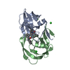
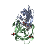
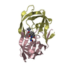
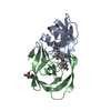
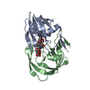
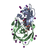
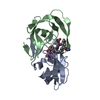
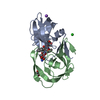
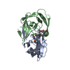
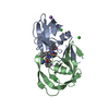
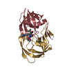
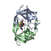
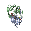
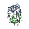
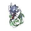
| Entry | Database: PDB / ID: 4flg | ||||||
|---|---|---|---|---|---|---|---|
| Title | HIV-1 protease mutant I47V complexed with reaction intermediate | ||||||
 Components Components |
| ||||||
 Keywords Keywords |  HYDROLASE / catalytic mechanism / drug resistance / aspartic protease HYDROLASE / catalytic mechanism / drug resistance / aspartic protease | ||||||
| Function / homology |  Function and homology information Function and homology informationRNA stem-loop binding / host cell / HIV-1 retropepsin / retroviral ribonuclease H / exoribonuclease H / exoribonuclease H activity / host multivesicular body / DNA integration / RNA-directed DNA polymerase / viral penetration into host nucleus ...RNA stem-loop binding / host cell / HIV-1 retropepsin / retroviral ribonuclease H / exoribonuclease H / exoribonuclease H activity / host multivesicular body / DNA integration / RNA-directed DNA polymerase / viral penetration into host nucleus / viral genome integration into host DNA / establishment of integrated proviral latency / RNA-directed DNA polymerase activity / RNA-DNA hybrid ribonuclease activity / Transferases; Transferring phosphorus-containing groups; Nucleotidyltransferases / viral nucleocapsid / DNA recombination / Hydrolases; Acting on ester bonds / DNA-directed DNA polymerase / aspartic-type endopeptidase activity / DNA-directed DNA polymerase activity / symbiont entry into host cell / symbiont-mediated suppression of host gene expression / lipid binding / host cell nucleus / host cell plasma membrane / virion membrane / structural molecule activity / proteolysis / DNA binding / zinc ion binding / membraneSimilarity search - Function | ||||||
| Biological species |   Human immunodeficiency virus 1 Human immunodeficiency virus 1 | ||||||
| Method | X-RAY DIFFRACTION / SYNCHROTRON / AB INITIO PHASING / Resolution: 1.31 Å | ||||||
 Authors Authors | Yu, X. / Shen, C.H. / Weber, I.T. | ||||||
 Citation Citation | Journal: Biochemistry / Year: 2012 Title: Capturing the Reaction Pathway in Near-Atomic-Resolution Crystal Structures of HIV-1 Protease. Authors: Shen, C.H. / Tie, Y. / Yu, X. / Wang, Y.F. / Kovalevsky, A.Y. / Harrison, R.W. / Weber, I.T. | ||||||
| History |
|
- Structure visualization
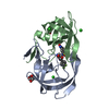
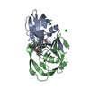
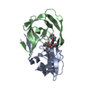
| Structure viewer | Molecule: MolmilJmol/JSmol |
|---|
- Downloads & links
-Download
| PDBx/mmCIF format | 4flg.cif.gz | 104.7 KB | Display | PDBx/mmCIF format |
|---|---|---|---|---|
| PDB format | pdb4flg.ent.gz | 80 KB | Display | PDB format |
| PDBx/mmJSON format | 4flg.json.gz | Tree view | PDBx/mmJSON format | |
| Others |  Other downloads Other downloads |
-Validation report
| Arichive directory | https://data.pdbj.org/pub/pdb/validation_reports/fl/4flgftp://data.pdbj.org/pub/pdb/validation_reports/fl/4flg | HTTPS FTP |
|---|
-Related structure data
| Related structure data |  4fl8C  4fm6C 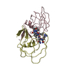 1fg6S C: citing same article ( S: Starting model for refinement |
|---|---|
| Similar structure data |


-Links
 PDBj
PDBj


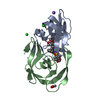
- Assembly
| Deposited unit | 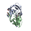
| ||||||||
|---|---|---|---|---|---|---|---|---|---|
| 1 |
| ||||||||
| Unit cell |
|
-Components
-Protein , 1 types, 2 molecules BA
| #1: Protein | / PR / Retropepsin Mass: 10726.649 Da / Num. of mol.: 2 / Mutation: Q7K, L33I, I47V, L63I, C67A, C95A Source method: isolated from a genetically manipulated source Source: (gene. exp.) Human immunodeficiency virus 1 / Production host:  Escherichia coli (E. coli) / References: UniProt: P03367, HIV-1 retropepsin Escherichia coli (E. coli) / References: UniProt: P03367, HIV-1 retropepsin |
|---|
-HIV-1 protease, ... , 3 types, 3 molecules CEF
| #2: Protein/peptide | Mass: 487.547 Da / Num. of mol.: 1 / Fragment: see remark 999 Source method: isolated from a genetically manipulated source Source: (gene. exp.) Human immunodeficiency virus 1 / Gene: gag-pol / Plasmid: pET11a / Production host: Escherichia coli (E. coli) / Strain (production host): BL21(DE3) / References: UniProt: P03367 |
|---|---|
| #3: Protein/peptide | Mass: 372.460 Da / Num. of mol.: 1 / Fragment: see remark 999 Source method: isolated from a genetically manipulated source Source: (gene. exp.) Human immunodeficiency virus 1 / Production host: Escherichia coli (E. coli) / References: UniProt: P03367 |
| #4: Protein/peptide | Mass: 373.445 Da / Num. of mol.: 1 / Fragment: see remark 999 Source method: isolated from a genetically manipulated source Source: (gene. exp.) Human immunodeficiency virus 1 / Production host: Escherichia coli (E. coli) / References: UniProt: P03367 |
-Non-polymers , 6 types, 193 molecules 


| #5: Chemical | ChemComp-GLU / Glutamic acid Type: L-peptide linking / Mass: 147.129 Da / Num. of mol.: 1 / Source method: obtained synthetically / Formula: C5H9NO4 Type: L-peptide linking / Mass: 147.129 Da / Num. of mol.: 1 / Source method: obtained synthetically / Formula: C5H9NO4 | ||||
|---|---|---|---|---|---|
| #6: Chemical | ChemComp-ILE / Isoleucine Type: L-peptide linking / Mass: 131.173 Da / Num. of mol.: 1 / Source method: obtained synthetically / Formula: C6H13NO2 Type: L-peptide linking / Mass: 131.173 Da / Num. of mol.: 1 / Source method: obtained synthetically / Formula: C6H13NO2 | ||||
| #7: Chemical | ChemComp-NA /  Mass: 22.990 Da / Num. of mol.: 1 / Source method: obtained synthetically / Formula: Na Mass: 22.990 Da / Num. of mol.: 1 / Source method: obtained synthetically / Formula: Na | ||||
| #8: Chemical | ChemComp-CL / Chloride Mass: 35.453 Da / Num. of mol.: 4 / Source method: obtained synthetically / Formula: Cl Mass: 35.453 Da / Num. of mol.: 4 / Source method: obtained synthetically / Formula: Cl#9: Chemical | Glycerol Mass: 92.094 Da / Num. of mol.: 2 / Source method: obtained synthetically / Formula: C3H8O3 Mass: 92.094 Da / Num. of mol.: 2 / Source method: obtained synthetically / Formula: C3H8O3#10: Water | ChemComp-HOH / | WaterMass: 18.015 Da / Num. of mol.: 184 / Source method: isolated from a natural source / Formula: H2O |
-Details
| Sequence details | AUTHOR STATES THAT CHAIN C, E AND F ARE SELF PROTEOLYTIC PRODUCT OF HIV-1 PROTEASE. SEQUENCE OF ...AUTHOR STATES THAT CHAIN C, E AND F ARE SELF PROTEOLYTI |
|---|
-Experimental details
-Experiment
| Experiment | Method: X-RAY DIFFRACTION / Number of used crystals: 1 |
|---|
- Sample preparation
| Crystal | Density Matthews: 2.53 Å3/Da / Density % sol: 51.34 % |
|---|---|
| Crystal grow | Temperature: 298 K / pH: 5 Details: 0.05 M sodium acetate buffer, 1.2 M sodium formate, and 2.5% PEG8000, pH 5.0, temperature 298K |
-Data collection
| Diffraction | Mean temperature: 100 K | |||||||||||||||||||||||||||||||||||||||||||||||||
|---|---|---|---|---|---|---|---|---|---|---|---|---|---|---|---|---|---|---|---|---|---|---|---|---|---|---|---|---|---|---|---|---|---|---|---|---|---|---|---|---|---|---|---|---|---|---|---|---|---|---|
| Diffraction source | Source: SYNCHROTRON / Site: APS  / Beamline: 22-ID / Wavelength: 0.8 Å / Beamline: 22-ID / Wavelength: 0.8 Å | |||||||||||||||||||||||||||||||||||||||||||||||||
| Detector | Type: MARMOSAIC 300 mm CCD / Detector: CCD / Date: Oct 16, 2008 | |||||||||||||||||||||||||||||||||||||||||||||||||
| Radiation | Monochromator: Si220 / Protocol: SINGLE WAVELENGTH / Monochromatic (M) / Laue (L): M / Scattering type: x-ray | |||||||||||||||||||||||||||||||||||||||||||||||||
| Radiation wavelength | Wavelength: 0.8 Å / Relative weight: 1 | |||||||||||||||||||||||||||||||||||||||||||||||||
| Reflection | Resolution: 1.31→50 Å / Num. all: 56095 / Num. obs: 53227 / % possible obs: 98.8 % / Observed criterion σ(F): 2 / Observed criterion σ(I): 2 | |||||||||||||||||||||||||||||||||||||||||||||||||
| Reflection shell |
|
- Processing
| Software |
| |||||||||||||||||||||||||||||||||
|---|---|---|---|---|---|---|---|---|---|---|---|---|---|---|---|---|---|---|---|---|---|---|---|---|---|---|---|---|---|---|---|---|---|---|
| Refinement | Method to determine structure: AB INITIO PHASING Starting model: 1FG6 Resolution: 1.31→10 Å / Num. parameters: 16870 / Num. restraintsaints: 21928 / Cross valid method: FREE R / σ(F): 0 / Stereochemistry target values: ENGH AND HUBER / Details: NO CUTOFF FOR ANISOTROPIC REFINEMENT
| |||||||||||||||||||||||||||||||||
| Refine analyze | Num. disordered residues: 25 / Occupancy sum hydrogen: 1641.88 / Occupancy sum non hydrogen: 1732.4 | |||||||||||||||||||||||||||||||||
| Refinement step | Cycle: LAST / Resolution: 1.31→10 Å
| |||||||||||||||||||||||||||||||||
| Refine LS restraints |
|
