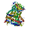
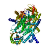
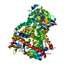
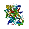
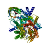
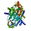
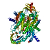
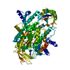
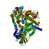
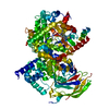
Entry Database : PDB / ID : 4fjyTitle Crystal structure of PI3K-gamma in complex with quinoline-indoline inhibitor 24f Phosphatidylinositol 4,5-bisphosphate 3-kinase catalytic subunit gamma isoform Keywords / / / / / / / / / Function / homology Function Domain/homology Component
/ / / / / / / / / / / / / / / / / / / / / / / / / / / / / / / / / / / / / / / / / / / / / / / / / / / / / / / / / / / / / / / / / / / / / / / / / / / / / / / / / / / / / / / / / / / / / / / / / / / / / / / / / / / / / / / / / / / / / / / / / / / / / / / Biological species Homo sapiens (human)Method / / Resolution : 2.9 Å Authors Whittington, D.A. / Tang, J. / Yakowec, P. Journal : J.Med.Chem. / Year : 2012Title : Discovery and in Vivo Evaluation of Dual PI3K-beta/delta inhibitorsAuthors: Gonzalez-Lopez de Turiso, F. / Shin, Y. / Brown, M. / Cardozo, M. / Chen, Y. / Fong, D. / Hao, X. / He, X. / Henne, K. / Hu, Y.L. / Johnson, M.G. / Kohn, T. / Lohman, J. / McBride, H.J. / ... Authors : Gonzalez-Lopez de Turiso, F. / Shin, Y. / Brown, M. / Cardozo, M. / Chen, Y. / Fong, D. / Hao, X. / He, X. / Henne, K. / Hu, Y.L. / Johnson, M.G. / Kohn, T. / Lohman, J. / McBride, H.J. / McGee, L.R. / Medina, J.C. / Metz, D. / Miner, K. / Mohn, D. / Pattaropong, V. / Seganish, J. / Simard, J.L. / Wannberg, S. / Whittington, D.A. / Yu, G. / Cushing, T.D. History Deposition Jun 12, 2012 Deposition site / Processing site Revision 1.0 Oct 24, 2012 Provider / Type Revision 1.1 Feb 28, 2024 Group / Database references / Derived calculationsCategory chem_comp_atom / chem_comp_bond ... chem_comp_atom / chem_comp_bond / database_2 / struct_ref_seq_dif / struct_site Item _database_2.pdbx_DOI / _database_2.pdbx_database_accession ... _database_2.pdbx_DOI / _database_2.pdbx_database_accession / _struct_ref_seq_dif.details / _struct_site.pdbx_auth_asym_id / _struct_site.pdbx_auth_comp_id / _struct_site.pdbx_auth_seq_id
Show all Show less
 Movie
Movie Controller

 Yorodumi
 Open data
 Basic information
 Components
 Keywords
 inflammation /
  Function and homology information
 Authors
 Citation
 Structure visualization
 Downloads & links
 Other downloads
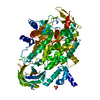


 PDBj
PDBj


 Assembly
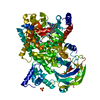


 Mass: 96.063 Da / Num. of mol.: 3 / Source method: obtained synthetically / Formula: SO4
 Mass: 468.565 Da / Num. of mol.: 1 / Source method: obtained synthetically / Formula: C29H29FN4O
 Mass: 18.015 Da / Num. of mol.: 13 / Source method: isolated from a natural source / Formula: H2O
 Sample preparation
 Processing